Review
. 2012 Feb;22(2):99-111.
doi: 10.1016/j.nmd.2011.10.009. Epub 2011 Nov 21.

Current status of the congenital myasthenic syndromes

Affiliations
Review

Current status of the congenital myasthenic syndromes

Andrew G Engel. Neuromuscul Disord. 2012 Feb.

Abstract

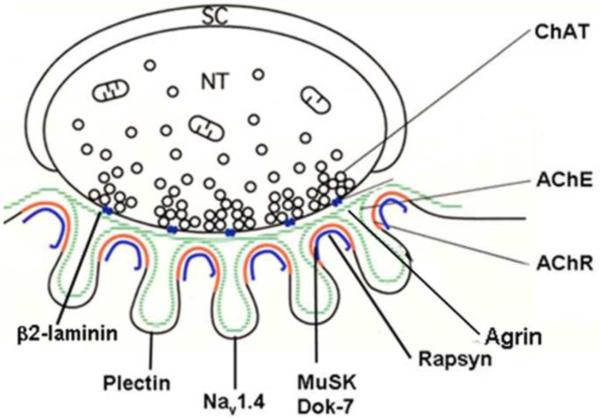
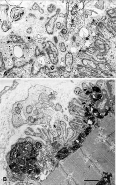
Congenital myasthenic syndromes (CMS) are heterogeneous disorders in which the safety margin of neuromuscular transmission is compromised by one or more specific mechanisms. Clinical, electrophysiologic, and morphologic studies have paved the way for detecting CMS-related mutations in proteins residing in the nerve terminal, the synaptic basal lamina, and in the postsynaptic region of the motor endplate. The disease proteins identified to date include choline acetyltransferase (ChAT), the endplate species of acetylcholinesterase (AChE), β2-laminin, the acetylcholine receptor (AChR), rapsyn, plectin, Na(v)1.4, the muscle specific protein kinase (MuSK), agrin, downstream of tyrosine kinase 7 (Dok-7), and glutamine-fructose-6-phosphate transaminase 1 (GFPT1). Myasthenic syndromes associated with centronuclear myopathies were recently recognized. Analysis of properties of expressed mutant proteins contributed to finding improved therapy for most CMS. Despite these advances, the molecular basis of some phenotypically characterized CMS remains elusive. Moreover, other types of CMS and disease genes likely exist and await discovery.


Figures

Fig. 1
Fig. 1
Schematic diagram of an EP with locations of presynaptic, synaptic and postsynaptic CMS disease proteins. Green line, synaptic basal lamina; red line, AChR on crests of the junctional folds; blue line, MuSK and Dok-7 closely associated with AChR. GFPT1, present in all tissues and potentially affecting multiple proteins, is not represented.
Fig. 2
Fig. 2
Schematic diagram showing domains of a ColQ strand and components of the asymmetric species of AChE. The indicated mutations appear in each ColQ domain.
Fig. 3
Fig. 3
Slow-channel syndrome. Repetitive compound muscle potentials (A) are generated by prolonged AChR channel opening events (B) that generate abnormally prolonged endplate currents (C). The slow-channel MEPC decays biexponentially owing to presence of both wild-type and mutant channels at the endplate. The αV249F mutation is in the second transmembrane domain of AChR. The prolonged endplate currents as well as leakiness of the receptor in the resting state result in Ca2+ overloading of the postsynaptic region (D and E) and in an EP myopathy (F). The slow-channel mutations occur in extracellular or transmembrane domains of each AChR subunit. Reproduced from Ref. 43, by permission.
Fig. 4
Fig. 4
Structural features of rapsyn deficient EPs. (A) Small cholinesterase reactive EP regions are dispersed over an extended length of the muscle fiber. These synaptic contacts differ from the compact pretzel shaped contacts at normal EPs. (B) and (C) Multiple small nerve terminals are apposed against a highly simplified postsynaptic regions with no (B) or few (C) junctional folds. In (B), note patchy distribution of AChR, visualized with peroxidase-labeled α-bungarotoxin, on the postsynaptic membrane. Bars 50 μm in (A) and 1 μm in (B) and (C). Reproduced from Ref. 71, by permission.
Fig. 5
Fig. 5
Dok-7 myasthenia. EPs with pre- and postsynaptic abnormalities. (A) EP region in shows marked degeneration of its junctional folds (asterisks). Schwann cell process (SC) is present amidst relics of the folds. A nerve sprout appears near the top. (B) A highly abnormal EP region devoid of nerve terminal. Some junctional folds are degenerating (asterisk). The subsynaptic sarcoplasm harbors large myeloid structures. A nerve sprouts surrounded by Schwann cell (SC) appears above the junction. Reproduced Ref. 3, by permission.

Similar articles

Cited by

References

    1. Engel AG, Lambert EH, Gomez MR. A new myasthenic syndrome with end-plate acetylcholinesterase deficiency, small nerve terminals, and reduced acetylcholine release. Ann Neurol. 1977;1:315–330. - PubMed
    1. Beeson D, Higuchi O, Palace J, et al. Dok-7 mutations underlie a neuromuscular junction synaptopathy. Science. 2006;313:1975–1978. - PubMed
    1. Selcen D, Milone M, Shen X-M, et al. Dok-7 myasthenia: phenotypic and molecular genetic studies in 16 patients. Ann Neurol. 2008;64:71–87. - PMC - PubMed
    1. Maselli RA, Ng JJ, Andreson JA, et al. Mutations in LAMB2 causing a severe form of synaptic congenital myasthenic syndrome. J Med Genet. 2009;46:203–208. - PMC - PubMed
    1. Liewluck T, Lovell TL, Bite AV, Engel AG. Sporadic centronuclear myopathy with muscle pseudohypertrophy, neutropenia, and necklace fibers due to a DNM2 mutation. Neuromuscul Disord. 2010;20:801–804. - PMC - PubMed

Publication types

MeSH terms

Substances