. 2012 Jan 2;287(1):748-756.
doi: 10.1074/jbc.M111.281295. Epub 2011 Nov 21.

Intra-membrane oligomerization and extra-membrane oligomerization of amyloid-β peptide are competing processes as a result of distinct patterns of motif interplay

Affiliations

Intra-membrane oligomerization and extra-membrane oligomerization of amyloid-β peptide are competing processes as a result of distinct patterns of motif interplay

Yi-Jiong Zhang et al. J Biol Chem. .

Abstract

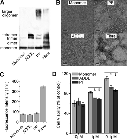
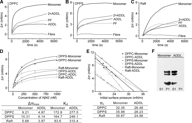
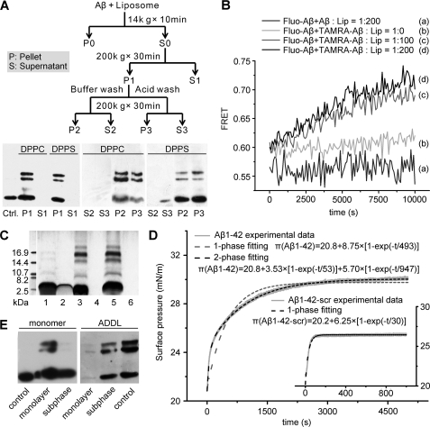
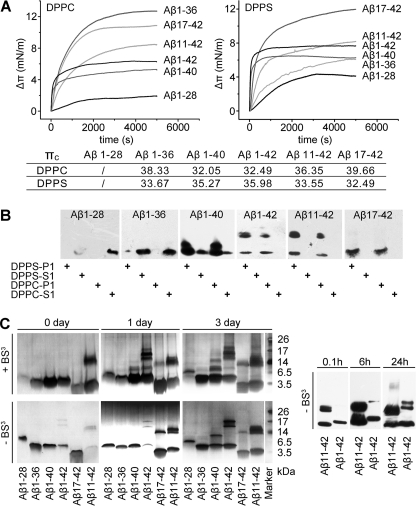
Soluble oligomers of amyloid-β peptide (Aβ) are emerging as the primary neurotoxic species in Alzheimer disease, however, whether the membrane is among their direct targets that mediate the downstream adverse effects remains elusive. Herein, we show that multiple soluble oligomeric Aβ preparations, including Aβ-derived diffusible ligand, protofibril, and zinc-induced Aβ oligomer, exhibit much weaker capability to insert into the membrane than Aβ monomer. Aβ monomers prefer incorporating into membrane rather than oligomerizing in solution, and such preference can be reversed by the aggregation-boosting factor, zinc ion. Further analyses indicate that the membrane-embedded oligomers of Aβ are derived from rapid assembly of inserted monomers but not due to the insertion of soluble Aβ oligomers. By comparing the behavior of a panel of Aβ truncation variants, we demonstrate that the intra- and extra-membrane oligomerization are mutually exclusive processes that proceed through distinct motif interplay, both of which require the action of amino acids 37-40/42 to overcome the auto-inhibitory interaction between amino acids 29-36 and the N-terminal portion albeit via different mechanisms. These results indicate that intra- and extra-membrane oligomerization of Aβ are competing processes and emphasize a critical regulation of membrane on the behavior of Aβ monomer and soluble oligomers, which may determine distinct neurotoxic mechanisms.


Figures

FIGURE 1.
FIGURE 1.
Characterization of Aβ samples in different assembly states. The characteristics of Aβ monomer, ADDL, protofibril (PF), and fiber were assessed by immunoblotting with 6E10 (A), EM observation (scale bars represent 50 nm) (B), and thioflavin T (ThT) fluorescence (C). Aβ monomer primarily migrated as a single band at ∼4.5 kDa in SDS-PAGE and was invisible under EM, whereas ADDL was characterized by bands corresponding to trimer/tetramer in SDS-PAGE and particle-like appearances with even size distribution (∼5 nm) under EM. There were additional high molecular weight bands in PF and fiber, and these two samples showed expected morphology. The absence of fiber in Aβ monomer, ADDL, and PF preparations were further verified by the comparable low ThT fluorescence as compared with that of mature fiber. D, cell viability of N2a cells treated with different sterile Aβ species for 48 h was assessed by 3-(4,5-dimethylthiazol-2-yl)-2,5-diphenyltetrazolium bromide assay. Results (n > 3) are given as mean ± S.E.; *, p < 0.05; **, p < 0.005. At low concentrations PF and ADDL are significantly more toxic than monomer sample in cell viability assay.
FIGURE 2.
FIGURE 2.
Solution-phase oligomerization impairs membrane insertion of Aβ. A–C, 600 nm Aβ monomer, ADDL (1200 nm for 2×ADDL), protofibril, or fibril was injected into the subphase beneath DPPC, DPPS or lipid raft-mimic monolayers with an initial surface pressure of 22 ± 1 mN/m, and the surface pressure increase (Δπ) − time curves were recorded. Raft-mimic monolayer was composed of DOPC, sphingomyelin, cholesterol, and GM1 ganglioside with a mole ratio of 32:32:31:5. Injection of Aβ monomer evoked an abrupt increase in surface pressure, whereas a slow and moderate increase was induced by addition of ADDL and PF. These indicate that Aβ monomer exhibited much stronger membrane insertion capability than ADDL and PF. Mature fiber was essentially unable to insert monolayer. D, surface pressure increases of lipid monolayers with an initial surface pressure of 22 ± 1 mN/m evoked by Aβ monomer or ADDL at the indicated concentrations. The data were fitted by the Hill equation (n = 1), and the corresponding parameters were listed in the inset table. These quantitative analyses indicate Aβ monomer possesses significantly higher affinity and maximal insertion capacity than ADDL. E, surface pressure change (Δπ) − initial surface pressure (πi) plots of Aβ interaction with monolayers composed of different lipids. The values of critical insertion pressure (πc) of Aβ for these monolayers are listed in the inset table. Because the πc of ADDL is lower than 30 mN/m, the physiological lateral pressure of cell membrane, this suggests that, unlike Aβ monomer, ADDL is unable to directly insert membrane bilayer. F, 1 μm Aβ monomer or ADDL was incubated with 200 μm DPPC liposomes for 2 h at room temperature. Aβ in the liposome-bound (P1) and supernatant fractions (S1; please refer to the scheme shown in Fig. 3A for the detailed preparation protocol) were obtained by ultracentrifugation at 200,000 × g for 30 min and probed by immunoblotting with 6E10. Aβ monomer was detected only in the liposome fraction while ADDL was exclusively detected in the solution fraction.
FIGURE 3.
FIGURE 3.
Membrane-associated Aβ monomers undergo rapid oligomerization. A, 1 μm Aβ monomer was incubated with 200 μm DPPC or DPPS liposomes for 2 h at room temperature and further processed as indicated in the scheme. Aβ in the liposome-bound (P: pellet) and supernatant fractions (S: supernatant) with or without acidic buffer treatment were detected by immunoblotting with 6E10. The Ctrl. lane is Aβ monomer, which underwent identical treatment in the absence of liposomes. Almost all the Aβ monomers were sedimented with liposomes, and acid treatment was unable to release liposome-associated Aβ. B, 0.5 μm fluorescein-labeled monomeric Aβ (donor) and 0.5 μm tetramethylrhodamine (TAMRA)-labeled monomeric Aβ (acceptor) were co-incubated with liposomes at the indicated peptide/lipid ratios, and FRET signal was collected and calculated as described under “Material and Methods.” The rapid increase in FRET signal indicates membrane-induced efficient oligomerization of Aβ. By contrast, no FRET signal could be detected when liposomes were absent or unlabeled peptide was used as acceptor. C, membrane-associated Aβ oligomers were prepared by reconstituting Aβ-lipid fusion mixture (1:200 molar ratio) or incubating Aβ monomer with the preformed liposomes (1:200 molar ratio). Samples were cross-linked with bis(sulfosuccinimidyl) suberate before silver-staining SDS-PAGE. Lanes 1 and 2 are Aβ monomer controls without lipid or liposomes. Lanes 3 and 4 are pellet and supernatant fractions of reconstituted Aβ-lipid fusion sample, respectively. Lanes 5 and 6 are pellet and supernatant fractions of liposome-Aβ incubation sample, respectively. It was evident that the two samples showed similar self-assembly patterns. D, exponential fitting of π-t plots of Aβ monomer insertion into DPPC monolayer. The experimental data of wild-type Aβ insertion deviates from one-phase process fitting but fits well to a two-phase process, suggesting that after insertion Aβ undergoes further conformational changes. As a comparison, the kinetics of scrambled Aβ fit better to a one-phase process. E, 600 nm Aβ monomer or ADDL was injected into the subphase beneath DPPC with a constant surface pressure of 28 mN/m. After 5000 s, the monolayer and subphase fractions were separated for subsequent immunoblotting with 6E10. The control lane is the Aβ monomer or ADDL at the same concentration incubating for 5000 s. The monolayer-inserted Aβ was largely oligomeric while Aβ in the subphase remained monomeric.
FIGURE 4.
FIGURE 4.
Intra- and extra-membrane oligomerization of Aβ are competing processes. A, the influence of zinc and calcium ions on Aβ oligomerization in solution was assessed by immunoblotting with 6E10 and 4G8 for detection of Aβ1–42 and Aβ17–42, respectively. 50 μm Zn2+ markedly accelerated the self-assembly of Aβ1–42 but not Aβ17–42, whereas Ca2+ showed little effect. Co-incubation of 100 μm EDTA abrogated the effects of Zn2+. B, the influence of zinc and calcium ions on Aβ1–42 or Aβ17–42 monomer insertion into DPPC monolayer. The arrow indicates the time of peptide injection. Zinc ion almost abrogated the monolayer insertion of Aβ1–42 irrespective of whether zinc was preincubated with Aβ1–42 or was just present during insertion. By contrast, zinc ion did not affect the insertion of Aβ17–42, a truncated Aβ variant without zinc binding site. Calcium ion showed little effect on Aβ1–42 insertion. Znmonolayer, injection of Zn2+ alone into the metal ion-free subphase; Aβ1–42monolayer + Zn, injection of Aβ1–42 alone into the subphase containing Zn2+; Aβ17–42monolayer + Zn, injection of Aβ17–42 alone into the subphase containing Zn2+; Aβ1–42 + Znmonolayer, injection of Aβ1–42 preincubated with Zn2+ into the metal ion-free subphase; Aβ17–42 + Znmonolayer, injection of Aβ17–42 preincubated with Zn2+ into the metal ion-free subphase; Aβ1–42monolayer + Ca, injection of Aβ1–42 alone into the subphase containing Ca2+; Aβ17–42monolayer + Ca, injection of Aβ17–42 alone into the subphase containing Ca2+; Aβ1–42 + Camonolayer, injection of Aβ1–42 preincubated with Ca2+ into the metal ion-free subphase; Aβ17–42 + Camonolayer, injection of Aβ17–42 preincubated with Ca2+ into the metal ion-free subphase; Zninserted Aβ1–42, injection of Zn2+ alone into the metal ion-free subphase beneath the monolayer with prior inserted Aβ1–42. C, 1 μm ADDL or Aβ monomer was incubated with DPPC liposomes for the indicated time and the liposome-bound (P1) and supernatant fractions (S1; please refer to the scheme shown in Fig. 3A for the detailed preparation protocol) were probed for Aβ signal by immunoblotting with 6E10. ADDL was incubated with DPPC liposomes for 24 h followed by 6E10 immunoblotting of liposome-bound (P2) and supernatant fractions (S2) after acidic buffer treatment (D) or EM observation (E). Prolonged incubation of ADDL with liposomes resulted in occurrence of liposome-associated Aβ oligomers, suggesting liposomes gradually shifts the equilibrium of monomers ↔ ADDL interconversion, thereby leading to the accumulation and oligomerization of Aβ in liposomes. The scale bar represents 100 nm. F, the proposed model depicting the distinct interactions of Aβ monomer and soluble oligomer with membrane. In the absence of aggregation-boosting factors (e.g. zinc ion) in solution, Aβ monomers preferentially insert membrane and undergoes rapid intra-membrane oligomerization. Appreciable solution-phase oligomerization may ensue only at elevated Aβ concentrations or with the co-existence of aggregation-boosting factors (e.g. zinc) to counteract the avid membrane interaction. Soluble oligomers are unable to insert membrane; rather they may function primarily by binding to high affinity cell surface receptors.
FIGURE 5.
FIGURE 5.
Intra- and extra-membrane assembly of Aβ proceed via distinct pathways. A, the Δπ-t curves of Aβ variant insertion into DPPC and DPPS monolayers with an initial surface pressure of 22 ± 1 mN/m. The values of πc were also indicated in the inset table. Except for Aβ1–28, all other peptides showed sufficient capacity to insert a physiological membrane. B, 1 μm Aβ variants were incubated with 200 μm DPPC or DPPS liposomes for 2 h at room temperature. The liposome-bound (P1) and supernatant fractions (S1; please refer to the scheme shown in Fig. 3A for the detailed preparation protocol) were probed by immunoblotting with 4G8. Almost no liposome association of Aβ1–28 and Aβ1–36 could be detected, whereas Aβ1–42, Aβ1–40, and Aβ11–42 showed a high level of membrane association and intra-membrane oligomerization. Despite strong membrane association, no intra-membrane oligomer of Aβ17–42 was observed. C, 100 μm Aβ variants were incubated in TBS at room temperature for the indicated times and analyzed by silver-staining SDS-PAGE (the left panel) with or without cross-linking, or by immunoblotting with 4G8 (the right panel) without prior cross-linking by bis(sulfosuccinimidyl) suberate (BS3). The aggregation patterns in silver-staining SDS-PAGE with or without cross-linking are very similar. At such a high concentration, all the Aβ variants aggregated albeit with different kinetics and capacity. Aβ1–28 is the most aggregative peptide showing no monomeric band in SDS-PAGE, whereas Aβ1–36 is the least aggregative peptide, the majority of which remained monomeric after a 3-day incubation.

References

    1. Citron M. (2010) Nat. Rev. Drug Discov. 9, 387–398 - PubMed
    1. Golde T. E., Schneider L. S., Koo E. H. (2011) Neuron 69, 203–213 - PMC - PubMed
    1. Hardy J., Selkoe D. J. (2002) Science 297, 353–356 - PubMed
    1. Hardy J. A., Higgins G. A. (1992) Science 256, 184–185 - PubMed
    1. Haass C., Selkoe D. J. (2007) Nat. Rev. Mol. Cell Biol. 8, 101–112 - PubMed

Publication types

Substances

LinkOut - more resources