. 2011 Dec;121(12):4712-21.
doi: 10.1172/JCI60417. Epub 2011 Nov 21.

EGFR as a therapeutic target for human, canine, and mouse ACTH-secreting pituitary adenomas

Affiliations

EGFR as a therapeutic target for human, canine, and mouse ACTH-secreting pituitary adenomas

Hidenori Fukuoka et al. J Clin Invest. 2011 Dec.

Abstract

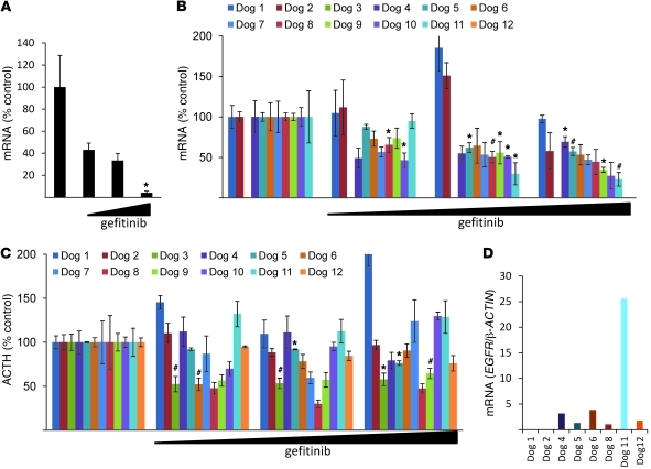
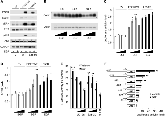
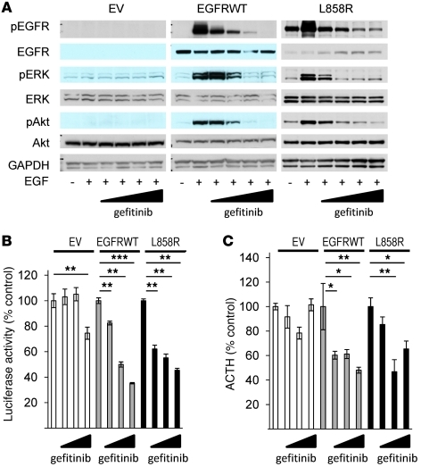
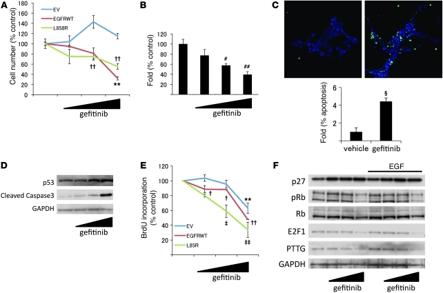
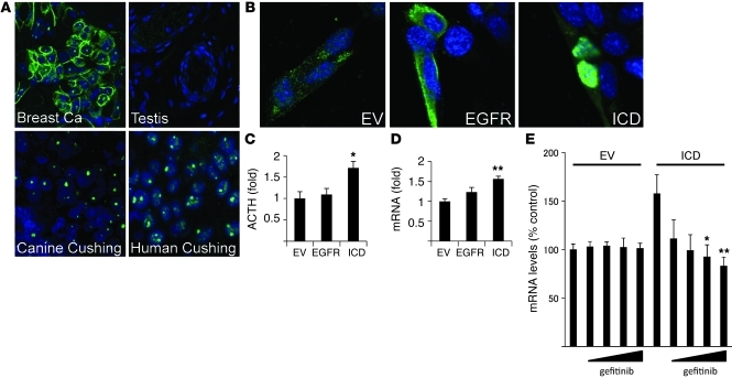
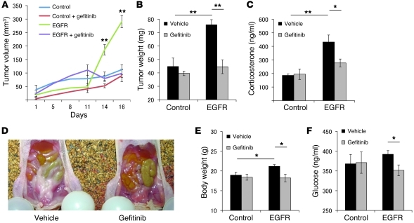
Cushing disease is a condition in which the pituitary gland releases excessive adrenocorticotropic hormone (ACTH) as a result of an adenoma arising from the ACTH-secreting cells in the anterior pituitary. ACTH-secreting pituitary adenomas lead to hypercortisolemia and cause significant morbidity and mortality. Pituitary-directed medications are mostly ineffective, and new treatment options are needed. As these tumors express EGFR, we tested whether EGFR might provide a therapeutic target for Cushing disease. Here, we show that in surgically resected human and canine corticotroph cultured tumors, blocking EGFR suppressed expression of proopiomelanocortin (POMC), the ACTH precursor. In mouse corticotroph EGFR transfectants, ACTH secretion was enhanced, and EGF increased Pomc promoter activity, an effect that was dependent on MAPK. Blocking EGFR activity with gefitinib, an EGFR tyrosine kinase inhibitor, attenuated Pomc expression, inhibited corticotroph tumor cell proliferation, and induced apoptosis. As predominantly nuclear EGFR expression was observed in canine and human corticotroph tumors, we preferentially targeted EGFR to mouse corticotroph cell nuclei, which resulted in higher Pomc expression and ACTH secretion, both of which were inhibited by gefitinib. In athymic nude mice, EGFR overexpression enhanced the growth of explanted ACTH-secreting tumors and further elevated serum corticosterone levels. Gefitinib treatment decreased both tumor size and corticosterone levels; it also reversed signs of hypercortisolemia, including elevated glucose levels and excess omental fat. These results indicate that inhibiting EGFR signaling may be a novel strategy for treating Cushing disease.


Figures

Figure 1
Figure 1. Effects of gefitinib on human and canine corticotroph adenoma cell cultures.
(A) After trans-sphenoidal surgical resection of human ACTH-secreting adenomas, tumor cells were dispersed, cultured, and treated with gefitinib (0.1–10 μM) for 24 hours, and human POMC expression levels were measured by real-time PCR. A representative experiment is shown (normalized by GAPDH). Each bar depicts 5 control replicates and 3 for each dose (0.1–10 μM). (BD) After trans-sphenoidal surgical resection of canine ACTH-secreting adenomas, tumor cells were dispersed, cultured, and treated with gefitinib (0.1–10 μM) for 24 hours. (B) Canine POMC expression levels were measured by real-time PCR. A representative experiment is shown (normalized by RPS5). (C) ACTH levels in culture media were measured using RIA. (D) Taqman PCR was performed using a specific canine EGFR probe and normalized by canine β-ACTIN. Values are mean ± SEM. *P < 0.05, #P < 0.01.
Figure 2
Figure 2. EGFR enhances Pomc mRNA expression and ACTH secretion via MAPK.
(A) EGFRWT, L858R, or EV AtT20 cells were treated with 5 nM EGF for 10 minutes, and Western blotting was performed. (B) EGFRWT cells were treated with EGF (0.5–50 nM) for the indicated times, and Northern blotting was performed. (C) EGFRWT, L858R, or EV cells were transfected with Pomc promoter and pRL-TK for 24 hours. After 6 hours of serum deprivation, cells were stimulated with EGF (0.5–50 nM) for 24 hours. (D) EGFRWT, L858R, or EV cells were treated with 5 nM EGF for 24 hours. ACTH secretion in the culture medium was determined by RIA (normalized for cell numbers). (E) EGFRWT cells were transfected with Pomc promoter and pRL-TK for 24 hours. After 6 hours of serum deprivation, cells were treated with U0126 (0.1–10 μM) or/and S31-201 (10–50 μM) for 45 minutes prior to induction with 5 nM EGF for 24 hours. (F) EGFRWT cells were transfected with Pomc promoter deletion mutants and pRL-TK for 24 hours. After 6 hours of serum deprivation, cells were stimulated with 5 nM EGF for 24 hours. Values are mean ± SEM. *P < 0.05, **P < 0.01, ***P < 0.05 vs. respective control (untreated EGFRWT or vehicle). Representative results are from triplicate samples in at least 2 independent experiments.
Figure 3
Figure 3. Gefitinib suppresses Pomc promoter activity and ACTH secretion.
(A) EGFRWT, L858R, or EV cells were treated with gefitinib (0.1–10 μM) for 45 minutes prior to induction with 5 nM EGF for 10 minutes, and Western blotting was performed. (B) EGFRWT, L858R, or EV cells were transfected with Pomc promoter and RSV–β-gal for 24 hours. After 6 hours of serum deprivation, cells were treated with gefitinib (0.1–10 μM) for 24 hours. (C) EGFRWT, L858R, or EV cells were treated with gefitinib (0.1–10 μM) for 24 hours, and ACTH secretion was determined by RIA (normalized for cell numbers). Values are mean ± SEM. *P < 0.05, **P < 0.01, ***P < 0.001. Representative results are from triplicate samples in at least 2 independent experiments.
Figure 4
Figure 4. Effects of gefitinib on corticotroph adenoma cell proliferation.
(A) EGFRWT, L858R, or EV cells were treated with gefitinib (0.1–10 μM) for 24 hours, and cells were counted. (B) EGFRWT cells were seeded (4,000 cells/well) for colony-forming assay, and gefitinib (0.1–10 μM) was added with serum-depleted media every third day. Colonies were counted from 5 randomly selected fields. (C) EGFRWT cells were treated with gefitinib (0.1–10 μM) for 48 hours, and TUNEL staining was performed. Original magnification, ×20. Apoptotic cells were counted from 5 randomly selected fields (normalized by DAPI-stained cell number). (D) EGFRWT cells were treated with gefitinib (0.1–10 μM) for 48 hours, and Western blotting was performed. (E) EGFRWT, L858R, or EV cells were treated with gefitinib (0.1–10 μM) for 24 hours, BrdU staining was performed, and incorporation was quantified using flow cytometry. (F) EGFRWT cells were treated with gefitinib (0.1–10 μM) for 24 hours with or without EGF (5 nM), and Western blotting was performed. Values are mean ± SEM. #P < 0.01, ##P < 0.01 vs. untreated control; §P < 0.001 vs. vehicle control; **P < 0.01 vs. EV control; P < 0.05, ††P < 0.01 vs. EGFRWT control; P < 0.05, ‡‡P < 0.01 vs. L858R control. Representative results are from triplicate samples in at least 2 independent experiments.
Figure 5
Figure 5. Nuclear EGFR localization in corticotroph adenomas.
(A) Confocal immunocytochemistry for EGFR was performed in human and canine corticotroph tumors. Human breast cancer tissue was used as a positive control, and human testis as negative control, for EGFR expression. Original magnification, ×100. (BE) AtT20 cells were transiently transfected with EGFR (pCMV/EGFR/myc/nuc), ICD (pCMV/EGFR-ICD/myc/nuc), or EV (pCMV/myc/nuc) for 24 hours. (B) Immunofluorescent chemistry was performed with anti-EGFR. Original magnification, ×100. (C) Media were changed to serum-depleted media and collected 24 hours later. ACTH levels in culture media were measured using RIA (normalized for cell numbers). (D) RNA was extracted, and mouse Pomc expression levels were measured by real-time PCR. A representative experiment is shown (normalized by Gapdh). (E) After gefitinib treatment for 24 hours, RNA was extracted, and mouse Pomc expression levels were measured by real-time PCR. A representative experiment is shown (normalized by Gapdh). Values are mean ± SEM. *P < 0.05, **P < 0.01, vs. EV. Representative results are from triplicate samples in at least 2 independent experiments.
Figure 6
Figure 6. Gefitinib attenuates EGFR tumor growth and hormone secretion in vivo.
EGFRWT (1 × 10⁶ cells/mouse, 0.1 ml with matrigel) or EV transfectants were injected s.c. in 7-week-old nude mice. 3 days after inoculation, mice with EGFR or control tumors received either vehicle (0.5% methylcellose and 0.5% tween80/PBS) or gefitinib (100 mg/kg) for 10 days. (A) On the indicated days after treatment, tumor volume was measured by caliper and calculated as in Methods. (B) After euthanasia, tumors were excised and weighed. (C) Serum corticosterone levels were measured by RIA. (D) Omental fat was more prominent in EGFR-injected than control mice. Murine body weight (E) and plasma glucose levels (F) were measured after euthanasia. Values are mean ± SEM. *P < 0.05; **P < 0.01.

Comment in

References

    1. Melmed S. Mechanisms for pituitary tumorigenesis: the plastic pituitary. J Clin Invest. 2003;112(11):1603–1618. - PMC - PubMed
    1. Newell-Price J, Bertagna X, Grossman AB, Nieman LK. Cushing’s syndrome. Lancet. 2006;367(9522):1605–1617. - PubMed
    1. Biller BM, et al. Treatment of adrenocorticotropin-dependent Cushing’s syndrome: a consensus statement. J Clin Endocrinol Metab. 2008;93(7):2454–2462. doi: 10.1210/jc.2007-2734. - DOI - PMC - PubMed
    1. Tritos NA, Biller BM, Swearingen B. Management of Cushing disease. Nat Rev Endocrinol. 2011;7(5):279–289. doi: 10.1038/nrendo.2011.12. - DOI - PubMed
    1. Jehle S, Walsh JE, Freda PU, Post KD. Selective use of bilateral inferior petrosal sinus sampling in patients with adrenocorticotropin-dependent Cushing’s syndrome prior to transsphenoidal surgery. J Clin Endocrinol Metab. 2008;93(12):4624–4632. doi: 10.1210/jc.2008-0979. - DOI - PMC - PubMed

Publication types

MeSH terms