. 2011 Dec;121(12):4850-60.
doi: 10.1172/JCI59261. Epub 2011 Nov 21.

Fate tracing of mature hepatocytes in mouse liver homeostasis and regeneration

Affiliations

Fate tracing of mature hepatocytes in mouse liver homeostasis and regeneration

Yann Malato et al. J Clin Invest. 2011 Dec.

Abstract

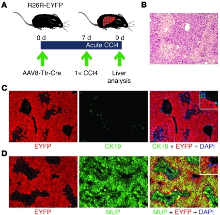
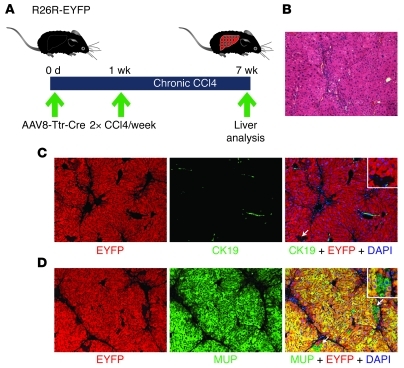
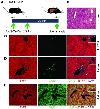
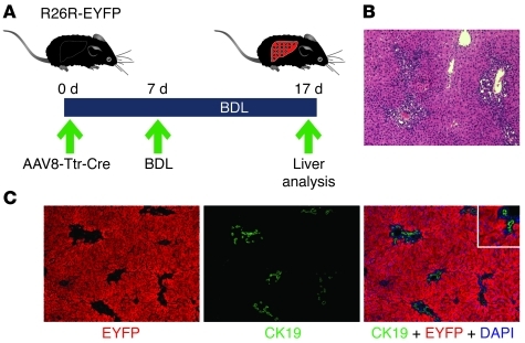
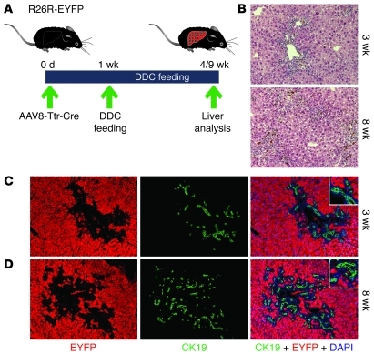
Recent evidence has contradicted the prevailing view that homeostasis and regeneration of the adult liver are mediated by self duplication of lineage-restricted hepatocytes and biliary epithelial cells. These new data suggest that liver progenitor cells do not function solely as a backup system in chronic liver injury; rather, they also produce hepatocytes after acute injury and are in fact the main source of new hepatocytes during normal hepatocyte turnover. In addition, other evidence suggests that hepatocytes are capable of lineage conversion, acting as precursors of biliary epithelial cells during biliary injury. To test these concepts, we generated a hepatocyte fate-tracing model based on timed and specific Cre recombinase expression and marker gene activation in all hepatocytes of adult Rosa26 reporter mice with an adenoassociated viral vector. We found that newly formed hepatocytes derived from preexisting hepatocytes in the normal liver and that liver progenitor cells contributed minimally to acute hepatocyte regeneration. Further, we found no evidence that biliary injury induced conversion of hepatocytes into biliary epithelial cells. These results therefore restore the previously prevailing paradigms of liver homeostasis and regeneration. In addition, our new vector system will be a valuable tool for timed, efficient, and specific loop out of floxed sequences in hepatocytes.


Figures

Figure 1
Figure 1. Rapid marker gene activation in all hepatocytes of adult R26R-EYFP and R26R mice injected with AAV8-Ttr-Cre.
(A) Results of AAV8-Ttr-Cre vector titration by dot blot and qPCR. The titer was approximately 4 × 10¹² viral genomes/ml in both dot blot and qPCR analysis (Supplemental Figure 1, A and B). Data represent mean ± SEM. (B) Dose-finding strategy. Livers were analyzed by immunostaining for EYFP 5 days after injection of different AAV8-Ttr-Cre doses into the tail veins of adult R26R-EYFP mice. (C) 1.5 × 10¹¹ viral genomes were not sufficient to activate EYFP expression (red) in all hepatocytes. 4 × 10¹¹ viral genomes activated EYFP expression in all hepatocytes. Nuclei were stained with DAPI (blue). (D) X-gal staining of livers of male and female R26R mice 48 hours after injection of 4 × 10¹¹ viral genomes shows β-gal expression (blue) in all hepatocytes. 15 liver sections from 3 mice were analyzed for each experiment. Original magnification, ×200.
Figure 2
Figure 2. AAV8-Ttr-Cre is not hepatotoxic.
(A) Toxicity testing. Blood and livers of adult R26R-EYFP mice were analyzed 3 and 5 days after injection of 4 × 10¹¹ viral genomes of AAV8-Ttr-Cre, AAV8-Ttr-control, or PBS, respectively. (B) H&E staining shows normal liver histologies 5 days after virus and PBS injection. Original magnification, ×100. 12 liver sections from 4 mice were analyzed per group. (C and D) Normal ALT and AST transaminase, direct bilirubin (DBIL), and albumin blood levels show that hepatocytes are not injured (C) and function normally (D) in AAV8-Ttr-Cre–injected mice. (E) qRT-PCR analysis shows that the inflammation-associated genes Tnfa and Il6 are not induced in livers of AAV8-Ttr-Cre–injected mice. 4 mice were analyzed per group. Data represent mean ± SEM.
Figure 3
Figure 3. Hepatocyte-specific activation of EYFP expression in adult R26R-EYFP mice injected with AAV8-Ttr-Cre.
Livers were coimmunostained for EYFP (red) and cell type–specific markers (green) 5 days after injection of 4 × 10¹¹ viral genomes of AAV8-Ttr-Cre. Double-positive cells appear yellow. (A) All MUP-positive cells are also EYFP positive, which confirms that AAV8-Ttr-Cre loops out floxed sequences in all hepatocytes of adult mice. (B) In Alb-Cre, R26R-EYFP control mice, not only all hepatocytes, but also all CK19-positive biliary cells, express EYFP. (C) All cells positive for CK19 are EYFP negative, which shows that AAV8-Ttr-Cre does not loop out floxed sequences in biliary epithelial cells and liver progenitor cells. (DF) All cells positive for F4/80 (D), desmin/α-SMA/GFAP (E), or isolectin B4 (“Lectin”) (F) are EYFP negative, which shows that AAV8-Ttr-Cre does not loop out floxed sequences in liver macrophages, stellate cells, or sinusoidal, portal vein, and central vein endothelial cells. Nuclei were stained with DAPI (blue). Original magnification, ×100, insets ×200 (A, C, and E); ×200, insets ×400 (B, D, and F). 20 liver sections from 4 mice were analyzed for each experiment.
Figure 4
Figure 4. Hepatocyte fate tracing in liver homeostasis.
(A) Livers of adult R26R-EYFP mice were analyzed 12 and 24 weeks after injection of 4 × 10¹¹ viral genomes of AAV8-Ttr-Cre. (B) H&E staining shows normal liver histologies. (C and D) Coimmunostaining for EYFP (red) and CK19 (green) shows normal hepatocyte plates and bile ducts 12 weeks (C) and 24 weeks (D) after AAV8-Ttr-Cre injection. No hepatocyte appears EYFP negative at both time points. (E and F) Coimmunostaining for EYFP (red) and MUP (green) confirms that all hepatocytes express EYFP 12 weeks (E) and 24 weeks (F) after AAV8-Ttr-Cre injection. Nuclei were stained with DAPI (blue). Original magnification, ×100, insets ×200. 15 liver sections from 3 mice were analyzed for each experiment.
Figure 5
Figure 5. Hepatocyte fate tracing after acute CCl4 intoxication.
(A) Adult R26R-EYFP mice were injected with 4 × 10¹¹ viral genomes of AAV8-Ttr-Cre and received a single dose of CCl4 7 days later. Livers were analyzed 2 days after CCl4 intoxication. (B) H&E staining shows pericentral hepatocyte necrosis. (C) Coimmunostaining for EYFP (red) and CK19 (green) confirms hepatocyte loss in pericentral areas, while periportal areas, including bile ducts, appear normal. (D) Coimmunostaining for EYFP (red) and MUP (green) fails to detect EYFP-negative hepatocytes emerging in periportal areas. Pericentral necrotic hepatocytes appear weakly MUP positive (Supplemental Figure 5C). Nuclei were stained with DAPI (blue). Original magnification, ×100, insets ×200. 20 liver sections from 4 mice were analyzed.
Figure 6
Figure 6. Hepatocyte fate tracing after chronic CCl4 intoxication.
(A) Adult R26R-EYFP mice were injected with 4 × 10¹¹ viral genomes of AAV8-Ttr-Cre and, starting 1 week later, received biweekly doses of CCl4 for 6 weeks. Livers were analyzed 3 days after the last CCl4 dose. (B) H&E staining shows liver remodeling, including bridging between periportal areas. (C) Coimmunostaining for EYFP (red) and CK19 (green) shows a cluster of EYFP-negative cells resembling hepatocytes in close proximity to a CK19-positive cell (white arrow). (D) Coimmunostaining for EYFP (red) and MUP (green) confirms the presence of clusters of EYFP-negative, MUP-positive hepatocytes (white arrows). Nuclei were stained with DAPI (blue). Original magnification, ×100, insets ×200. 15 liver sections from 3 mice were analyzed.
Figure 7
Figure 7. Hepatocyte fate tracing after 2/3 PH.
(A) Adult R26R-EYFP mice were injected with 4 × 10¹¹ viral genomes of AAV8-Ttr-Cre and underwent 2/3 PH 7 days later. Livers were analyzed 21 days after 2/3 PH. Liver lobes removed by 2/3 PH were used as controls. (B) H&E staining shows restoration of normal liver histology 21 days after 2/3 PH. (C and D) Coimmunostaining for EYFP (red) and CK19 (green) shows that all hepatocytes expressed EYFP before 2/3 PH (C) but that clusters of EYFP-negative cells resembling hepatocytes are present in periportal areas after 2/3 PH (D, white arrows). (E) Coimmunostaining for EYFP (red) and MUP (green) confirms the presence of single cells and clusters of EYFP-negative, MUP-positive hepatocytes (white arrows). Nuclei were stained with DAPI (blue). Original magnification, ×100, insets ×200. 20 liver sections from 4 mice were analyzed.
Figure 8
Figure 8. Hepatocyte fate tracing after BDL.
(A) Adult R26R-EYFP mice were injected with 4 × 10¹¹ viral genomes of AAV8-Ttr-Cre and underwent BDL 7 days later. Livers were analyzed 10 days after BDL. (B) H&E staining shows extensive ductular reactions. (C) Coimmunostaining for EYFP (red) and CK19 (green) shows no double-positive cells. Nuclei were stained with DAPI (blue). Original magnification, ×100, insets ×200. 25 liver sections from 5 mice were analyzed.
Figure 9
Figure 9. Hepatocyte fate tracing after DDC feeding.
(A) Adult R26R-EYFP mice were injected with 4 × 10¹¹ viral genomes of AAV8-Ttr-Cre, and DDC feeding began 1 week later. Livers were analyzed after 3 or 8 weeks of DDC feeding. (B) H&E staining shows ductular reactions expanding with time of DDC feeding. (C and D) Coimmunostaining for EYFP (red) and CK19 (green) shows no double-positive cells after 3 (C) or 8 (D) weeks of DDC feeding. Nuclei were stained with DAPI (blue). Original magnification, ×100, insets ×200. 20 liver sections from 4 mice were analyzed for each experiment.

Comment in

References

    1. Fausto N. Liver regeneration and repair: hepato­cytes, progenitor cells, and stem cells. Hepatology. 2004;39(6):1477–1487. doi: 10.1002/hep.20214. - DOI - PubMed
    1. Fausto N, Campbell JS, Riehle KJ. Liver regeneration. Hepatology. 2006;43(2 Suppl 1):S45–53. doi: 10.1002/hep.20969. - DOI - PubMed
    1. Michalopoulos GK. Liver regeneration. J Cell Physiol. 2007;213(2):286–300. doi: 10.1002/jcp.21172. - DOI - PMC - PubMed
    1. Duncan AW, Dorrell C, Grompe M. Stem cells and liver regeneration. Gastroenterology. 2009;137(2):466–481. doi: 10.1053/j.gastro.2009.05.044. - DOI - PMC - PubMed
    1. Michalopoulos GK. Liver regeneration: alternative epithelial pathways. Int J Biochem Cell Biol. 2011;43(2):173–179. doi: 10.1016/j.biocel.2009.09.014. - DOI - PMC - PubMed

Publication types

MeSH terms