. 2012 Jul 26;31(30):3505-15.
doi: 10.1038/onc.2011.518. Epub 2011 Nov 21.

A recombinant decoy comprising EGFR and ErbB-4 inhibits tumor growth and metastasis

Affiliations

A recombinant decoy comprising EGFR and ErbB-4 inhibits tumor growth and metastasis

M Lindzen et al. Oncogene. .

Abstract

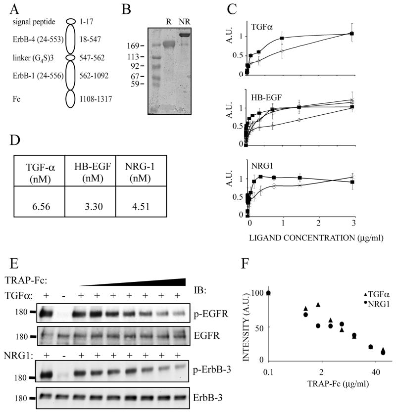
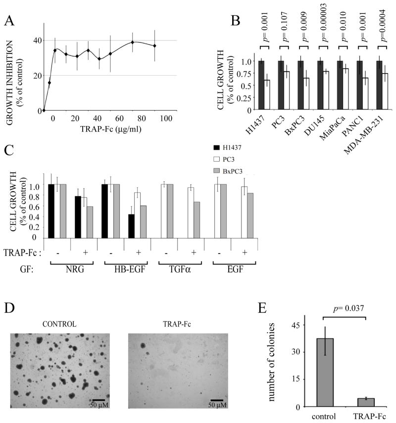
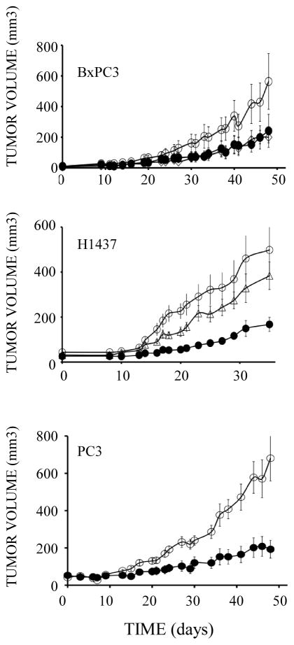
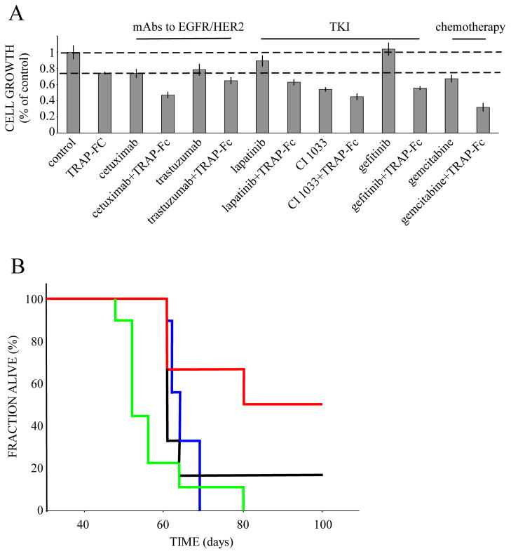
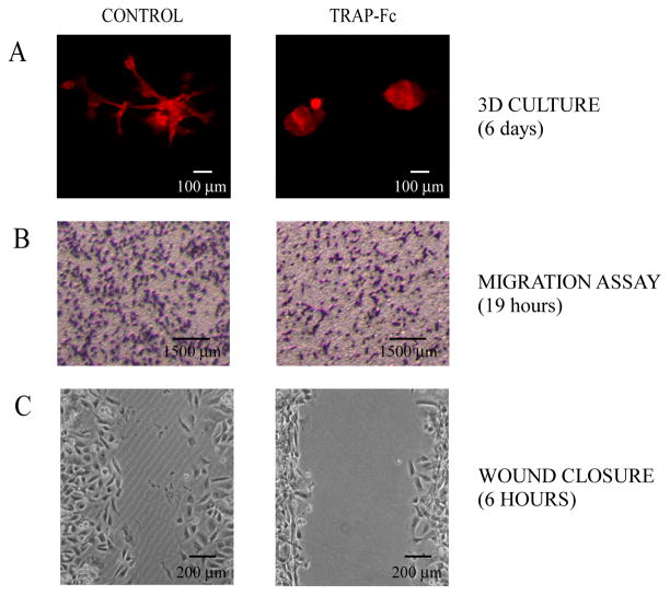
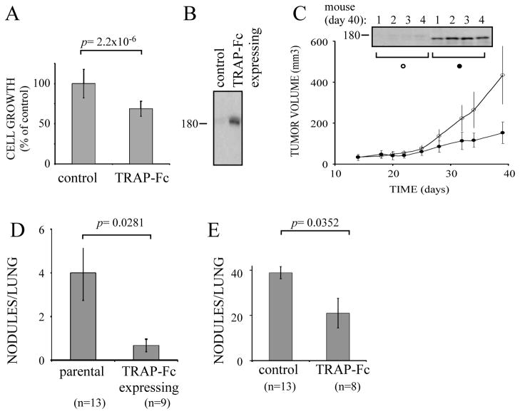
Epidermal growth factor (EGF)-like growth factors control tumor progression as well as evasion from the toxic effects of chemotherapy. Accordingly, antibodies targeting the cognate receptors, such as EGFR/ErbB-1 and the co-receptor HER2/ErbB-2, are widely used to treat cancer patients, but agents that target the EGF-like growth factors are not available. To circumvent the existence of 11 distinct ErbB ligands, we constructed a soluble fusion protein (hereinafter: TRAP-Fc) comprising truncated extracellular domains of EGFR/ErbB-1 and ErbB-4. The recombinant TRAP-Fc retained high-affinity ligand binding to EGF-like growth factors and partially inhibited growth of a variety of cultured tumor cells. Consistently, TRAP-Fc displayed an inhibitory effect in xenograft models of human cancer, as well as synergy with chemotherapy. Additionally, TRAP-Fc inhibited invasive growth of mammary tumor cells and reduced their metastatic seeding in the lungs of animals. Taken together, the activities displayed by TRAP-Fc reinforce critical roles of EGF-like growth factors in tumor progression, and they warrant further tests of TRAP-Fc in preclinical models.


Figures

Figure 1
Figure 1. Construction, expression and functional tests of TRAP-Fc
(A) A scheme of the recombinant TRAP-Fc protein, including a signal peptide, the three N-terminal extracellular sub-domains of ErbB-4 called LI (domain I), SI (domain II), LII (domain III) and a portion of SII (domain IV), a linker, followed by the corresponding portion of ErbB-1 linked to human immunoglobulin lambda’s Fc portion. Residue numbers corresponding to ErbB-4 and ErbB-1 appear in parentheses. Other numbers refer to the TRAP’s full sequence. (B) Coomassie blue staining of an acrylamide gel showing the purified TRAP-Fc protein (2μg) following electrophoresis under non-reducing (NR) or reducing (R) conditions. (C) ELISA plates were coated with IgB proteins or with TRAP-Fc (each at 8 μg/ml). Ligand binding and detection using streptavidin-HRP were performed as detailed under Methods. TRAP-Fc, full squares; IgB-1, diamonds; IgB-3, empty squares and IgB-4, crosses. (D) Solutions containing the TRAP-Fc (1–100 nM) were passed over surfaces coated with the indicated ligands, to derive the indicated dissociation constants using plasmon resonance measurements. (E) HeLa (upper panel) and T47D cells (lower panel) were incubated for 10 minutes with the indicated ligands (5 ng/ml) and increasing concentrations of TRAP-Fc (0, 0.8, 1.6, 3.2, 6, 12, 30, 60 μg/ml). Cleared cell extracts were immunoblotted (IB) with the indicated antibodies, including antibodies specific to the tyrosine phosphorylated forms of ErbB-1/EGFR (Tyr-1068) or ErbB-3 (Tyr-1289). (F) Densitometric analyses corresponding to the immunoblots presented in E.
Figure 2
Figure 2. The TRAP-Fc inhibits proliferation of human tumor cells
(A) BxPC3 pancreatic tumor cells (2×10⁴) were incubated with increasing concentrations of TRAP-Fc. Cell proliferation was determined in hexaplicates after 5 days, using the MTT assay. Averages ± S.D. (bars) are indicated. (B) The indicated cancer cells (2×10⁴) were incubated for 5 days with TRAP-Fc (20 μg/ml) and cell proliferation was determined as in A. (C) H1437 lung tumor cells, PC3 prostate tumor cells and BxPC3 pancreatic tumor cells (each at 2×10⁴ cells per well) were incubated with the indicated ligands (5 ng/ml), along with TRAP-Fc (20 μg/ml), and cell proliferation assayed as in A. (D and E) H1437 cells were seeded in agarose (top layer: 0.3% agar; bottom layer: 0.6% agar) in 6-well plates (1×10⁴ cells per well) and overlaid with medium. TRAP-Fc was added to both the soft agar and to the medium (at 100 μg/ml). Photographs were captured and colony numbers were determined in five randomly chosen high-powered fields. The data represent mean±S.D. The data sets were analyzed for statistically significant differences by using the two-tailed Student t test.
Figure 3
Figure 3. TRAP-Fc inhibits tumorigenic growth of human cancer cells in mice
Female nude mice (6 week old) were inoculated subcutaneously with the indicated human tumor cells (2×10⁶ per animal). Once tumors became palpable, mice were randomized into three groups and injected intraperitoneally with TRAP-Fc (100 μg; closed circles), or with a combination of anti-TGFα and anti HB-EGF mAbs (each at 250 μg/mouse, open triangle; BxPC3 and H1437 cells only). Mice bearing BxPC3 xenografts were injected on days: 9, 16, 20, 23, 26, 30, 33, 37, 40 and 44. Both the control and the treatment group included 8 mice. Note: two mice of the treatment group completely lost their tumors and were not included in the results. The mAb group included 3 mice. Mice bearing H1437 xerografts were injected on days 6, 10, 14, 18 and 21. The control group included 15 mice, the TRAP-Fc-treated group included 7 mice and the mAb group included 11 mice. Mice bearing PC3 xenografts were injected on days 1, 3, 6, 9, 13, 17, 20, 23, 27 and 30. The control group included 11 mice and the TRAP-Fc-treated group included 10 mice (two of them lost their tumors and are not represented).
Figure 4
Figure 4. Combinations of TRAP-Fc and therapeutic agents effectively inhibit growth of human pancreatic cancer cells in vitro and extend survival of tumor-bearing mice
(A) BxPC3 pancreatic tumor cells (2×10⁴) were incubated with TRAP-Fc (30 μg/ml), either alone or in combination with cetuximab (20 μg/ml), trastuzumab (20 μg/ml), lapatinib (0.05 nM), erlotinib (0.2 nM), CI-1033 (0.5 nM), AG1478 (0.2 nM), gefitinib (0.004 nM), or gemcitabine (at 0.5 ng/ml). Cellular proliferation was measured in hexaplicates after 5 days, using the MTT assay. The experiment was repeated twice. (B) Female nude mice (6-week old) were inoculated subcutaneously with BxPC3 pancreatic cancer cells (2×10⁶). Once tumors became palpable, mice were randomized into four groups. The control group (9 mice; blue) is shown. The TRAP-Fc-treated group (6 mice; black line) was injected intraperitoneally with TRAP-Fc (100 μg). A third group (9 mice) was treated with gemcitabine (intraperitoneal injection, 25 mg/kg; green line), and a fourth group (6 mice; red) was treated with a combination of gemcitabine and TRAP-Fc. Mice were injected with TRAP-Fc on days: 19, 28, 32, 35, 39, 42, and 45. Gemcitabine injections were given on the same days and repeated three more times on days: 49, 52 and 55. Body weights were measured once a week, but no consistent differences were observed (data not shown).
Figure 5
Figure 5. TRAP-Fc inhibits invasive growth of MDA-MB-231 mammary tumor cells in vitro
(A) Eight-chambered plates were coated with an extracellular matrix (Matrigel). MDA-MB-231 cells (2,000 cells/well) were mixed with medium containing Matrigel and then added to the chambers. Cells were incubated without or with TRAP-Fc (30 μg/ml), and phase contrast photomicrographs were captured six days later. (B) MDA-MB-231 cells (1.5×10⁵) were incubated in Transwell chambers in the absence or presence of TRAP-Fc (30 μg/ml). Cell migration across the filter separating the two compartments was measured 19 hours later and representative photographs of the lower faces of the filters were taken. Migration was normalized to the input number of cells. (C) MDA-MB-231 cells were plated on wound-healing inserts. Twenty-four hours later, plugs were removed, cells were treated with TRAP-Fc (100 μg/ml) and allowed to migrate. Snapshots captured after 6 hours are presented.
Figure 6
Figure 6. TRAP-Fc inhibits lung metastasis of MDA-MB-231 mammary cancer cell
(A) MDA-MB-231 cells (2×10⁴) were treated with TRAP-Fc (30 μg/ml) and proliferation was determined in hexaplicates after 5 days, using the MTT assay. (B) Conditioned media collected from parental cells and from MDA-MB-231 cells stably expressing the TRAP-Fc protein were immunoblotted (IB) with an anti-EGFR antibody. (C) Parental MDA-MB-231 and TRAP-Fc-expressing cells (2.5×10⁶) were inoculated into the mammary fat pad of scid mice (6 and 7 mice, respectively). Tumor size was monitored twice a week. Average tumor sizes and standard deviations (bars) are shown. In the end of the experiment (day 40) tumors were removed from 4 mice of each group, homogenized, electrophoresed and immunoblotted with an anti-EGFR antibody. Representative blots are shown. (D) Parental MDA-MB-231 and TRAP-Fc-expressing cells (2.5×10⁶) were injected into the tail vein of scid mice. Nodules in the lungs were counted at day 60 and their averages presented. The control group included 13 mice, and the TRAP-expressing group included 9 mice. (E) MDA-MB-231 cells (1.5×10⁵) were injected into the tail vein of scid mice. TRAP-Fc (100 μg per injection) was injected intraperitoneally (8 mice) on days 1, 3, 6 and 9. Nodules in the lungs were counted at day 60. The control group included 13 mice.

References

    1. Aboud-Pirak E, Hurwitz E, Pirak ME, Bellot F, Schlessinger J, Sela M. Efficacy of antibodies to epidermal growth factor receptor against KB carcinoma in vitro and in nude mice. J Natl Cancer Inst. 1988;80:1605–11. - PubMed
    1. Adams TE, Koziolek EJ, Hoyne PH, Bentley JD, Lu L, Lovrecz G, et al. A truncated soluble epidermal growth factor receptor-Fc fusion ligand trap displays anti-tumour activity in vivo. Growth Factors. 2009;27:141–54. - PubMed
    1. Atlas E, Cardillo M, Mehmi I, Zahedkargaran H, Tang C, Lupu R. Heregulin is sufficient for the promotion of tumorigenicity and metastasis of breast cancer cells in vivo. Mol Cancer Res. 2003;1:165–75. - PubMed
    1. Barozzi C, Ravaioli M, D’Errico A, Grazi GL, Poggioli G, Cavrini G, et al. Relevance of biologic markers in colorectal carcinoma: a comparative study of a broad panel. Cancer. 2002;94:647–57. - PubMed
    1. Baselga J. Targeting tyrosine kinases in cancer: the second wave. Science. 2006;312:1175–8. - PubMed

Publication types

MeSH terms