. 2012 Mar 27;18(1):201-8.
doi: 10.2119/molmed.2011.00282.

Interferon regulatory factor-1 regulates the autophagic response in LPS-stimulated macrophages through nitric oxide

Affiliations

Interferon regulatory factor-1 regulates the autophagic response in LPS-stimulated macrophages through nitric oxide

Lemeng Zhang et al. Mol Med. .

Abstract

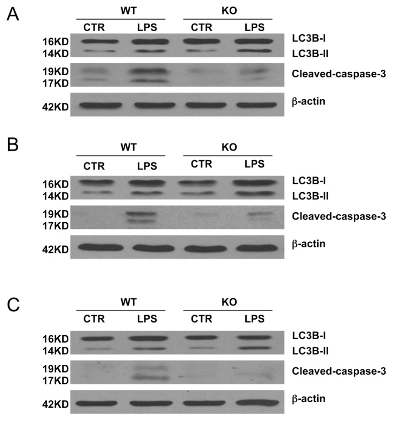
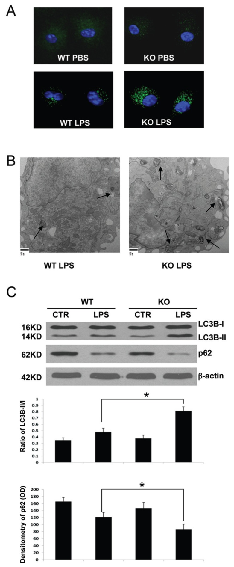
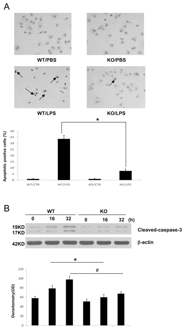
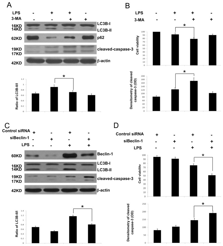
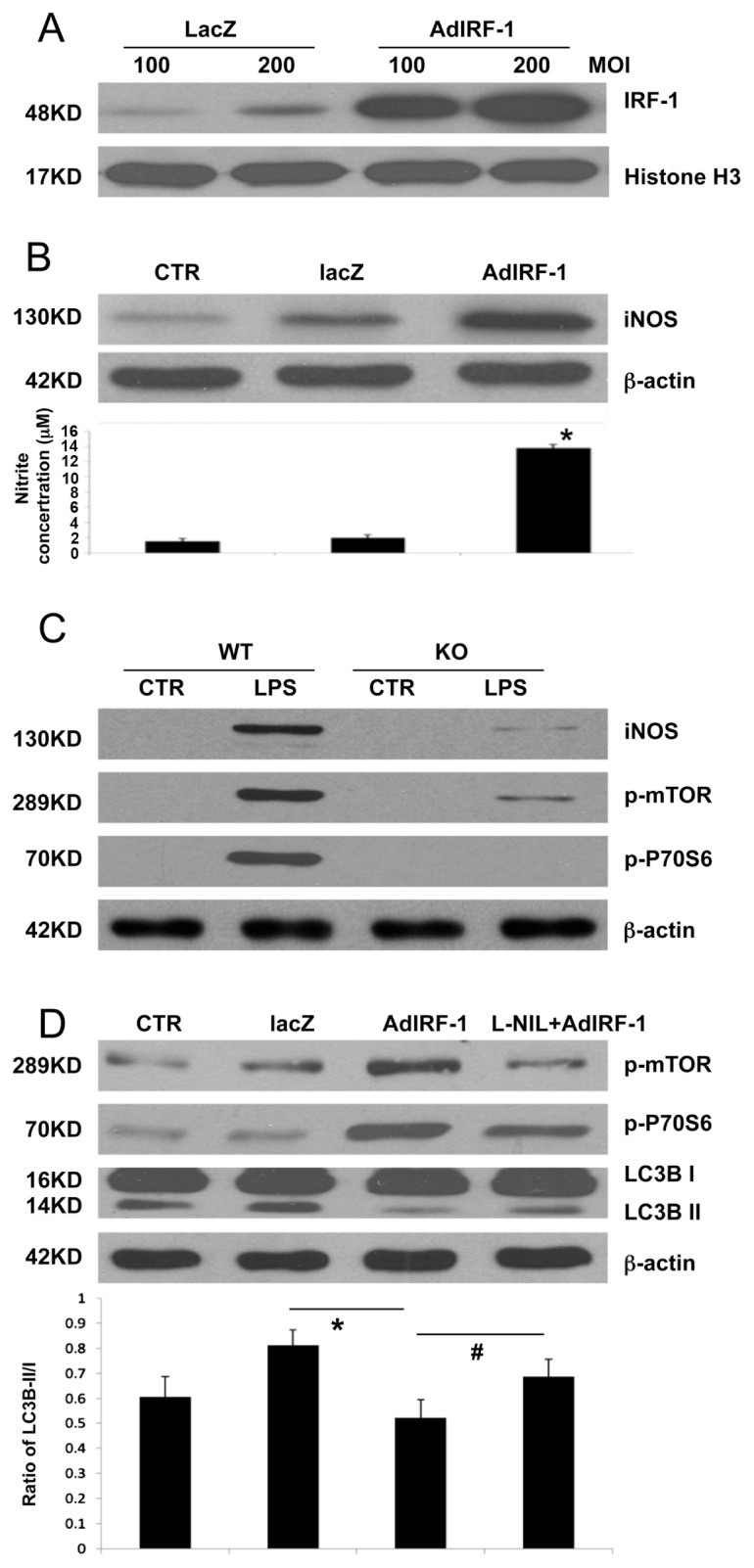
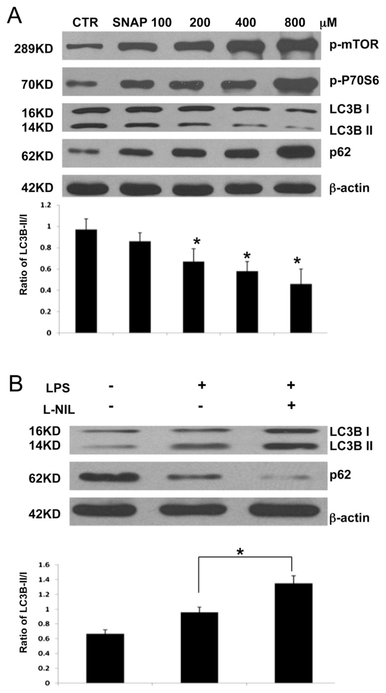
The pathogenesis of sepsis is complex and, unfortunately, poorly understood. The cellular process of autophagy is believed to play a protective role in sepsis; however, the mechanisms responsible for its regulation in this setting are ill defined. In the present study, interferon regulatory factor 1 (IRF-1) was found to regulate the autophagic response in lipopolysaccharide (LPS)-stimulated macrophages. In vivo, tissue macrophages obtained from LPS-stimulated IRF-1 knockout (KO) mice demonstrated increased autophagy and decreased apoptosis compared to those isolated from IRF-1 wild-type (WT) mice. In vitro, LPS-stimulated peritoneal macrophages obtained from IRF-1 KO mice experienced increased autophagy and decreased apoptosis. IRF-1 mediates the inhibition of autophagy by modulating the activation of the mammalian target of rapamycin (mTOR). LPS induced the activation of mTOR in WT peritoneal macrophages, but not in IRF-1 KO macrophages. In contrast, overexpression of IRF-1 alone increased the activation of mTOR and consequently decreased autophagic flux. Furthermore, the inhibitory effects of IRF-1 mTOR activity were mediated by nitric oxide (NO). Therefore, we propose a novel role for IRF-1 and NO in the regulation of macrophage autophagy during LPS stimulation in which IRF-1/NO inhibits autophagy through mTOR activation.


Figures

Figure 1
Figure 1
Macrophages from IRF-1 KO mice exhibit decreased apoptosis and increased autophagy during endotoxemia. (A) Following PBS or LPS (20 mg/kg) administration for 16 h, splenic macrophages were isolated from WT and IRF-1 KO mice and analyzed by Western blot for LC3B I/II and cleaved caspase-3. (B) Following PBS or LPS (20 mg/kg) administration for 16 h, alveolar macrophages were isolated from WT and IRF-1 KO mice and analyzed by Western blot for LC3B I/II and cleaved caspase-3. (C) Following PBS or LPS (20 mg/kg) administration for 16 h, liver Kupffer cells were isolated from WT and IRF-1 KO mice and analyzed by Western blot for LC3B I/II and cleaved caspase-3. Results are representative of three separate independent experiments.
Figure 2
Figure 2
Peritoneal macrophages from IRF-1 KO mice demonstrate increased autophagy in response to LPS stimulation. (A) Peritoneal macrophages from C57BL/6 and IRF-1 KO mice were isolated and treated with/without LPS (1 μg/mL) for 16 h; Immunofluorescent confocal microscopy was then performed (magnification 600×). (B) Peritoneal macrophages were isolated from WT and IRF-1 KO mice and stimulated with LPS (1 μg/mL) for 16 h and analyzed by TEM for autophagosomes and autolysosomes (magnification 25,000×). Scale bars = 500 nm. (C) Peritoneal macrophages were isolated from WT and IRF-1 KO mice and stimulated with LPS (1 μg/mL) for 16 h and were analyzed by Western blot for LC3B I/II and p62. *P < 0.05; results representative of three separate independent experiments.
Figure 3
Figure 3
Peritoneal macrophages from IRF-1 KO mice demonstrate decreased apoptosis in response to LPS stimulation. Peritoneal macrophages were isolated from WT and KO mice and were treated with/without LPS (1 μg/mL) stimulation for 16 h or 32h and were analyzed by (A) TUNEL staining or (B) cleaved caspase-3 by Western blot. *,#P < 0.05; results representative of three separate independent experiments.
Figure 4
Figure 4
Decreased autophagic flux in IRF-1 WT macrophages leads to apoptotic cell death. (A) RAW264.7 cells were pretreated with the autophagy inhibitor 3-MA (5 mmol/L) for 1 h prior to LPS (500 ng/mL) stimulation. Following LPS stimulation for 16 h, cells were analyzed by Western blot for LC3B I/II, p62 and caspase-3 cleavage. (B) Cell viability was analyzed by MTT assay. (C) RAW 264.7 cells were transfected with siBeclin-1 for 24 h prior to LPS (500 ng/mL) stimulation. Following LPS stimulation for 16 h, cells were analyzed by western blot for LC3B I/II, p62 and caspase-3 cleavage. (D) Cell viability was analyzed by MTT assay. *P < 0.05; results representative of three separate independent experiments.
Figure 5
Figure 5
IRF-1 inhibits autophagy through iNOS/mTOR/P70S6 in vitro. (A) AdIRF-1 infection induced IRF-1 overproduction. AdLacZ and AdIRF-1 infected RAW 264.7 cells for 24 h at either an MOI of 100 or 200 and Western blot analysis for IRF-1 expression was performed. (B) RAW cells were infected with AdIRF-1 at an MOI of 200 for 24 h; cells were then harvested for iNOS expression. Griess assay confirmed NO production following AdIRF-1 infection. (C) Peritoneal macrophages were isolated from WT and IRF-1 KO mice and stimulated with LPS (1 μg/mL) for 16 h and analyzed by Western blot for iNOS, p-mTOR and p-P70S6. (D) RAW cells were infected with AdIRF-1 at an MOI of 200 for 24 h and were treated with L-NIL (200 μmol/L) for 16 h. Cells were then harvested for Western blot of p-mTOR, p-P70S6, LC3B I/II and P62. *,#P < 0.05. Results representative of three separate independent experiments.
Figure 6
Figure 6
NO/iNOS mimics the effects seen in IRF-1 overexpression on autophagy in RAW264.7 cells. (A) Raw cells were pre-treated with serum-free media for 12 h and then stimulated with a different dose of SNAP, an NO donor for 16 h. Cells were harvested for western blot analysis for p-mTOR, p-P70S6, LC3B I/II and P62. *P < 0.05; Results representative of three separate independent experiments. (B) RAW 264.7 cells were pretreated with L-NIL (200 μmol/L) for 1h and then stimulated with LPS (500 ng/mL) for 16 h. Cells were harvested for Western blot analysis for LC3B and p62. *P < 0.05; results representative of three separate independent experiments.

Similar articles

Cited by

References

    1. Anderson RN, Smith BL. Deaths: leading causes for 2002. Natl Vital Stat Rep. 2005;53:1–89. - PubMed
    1. Chopra M, Sharma AC. Distinct cardiodynamic and molecular characteristics during early and late stages of sepsis-induced myocardial dysfunction. Life Sci. 2007;81:306–16. - PMC - PubMed
    1. Xiao H, Siddiqui J, Remick DG. Mechanisms of mortality in early and late sepsis. Infect Immun. 2006;74:5227–35. - PMC - PubMed
    1. Rubinsztein DC, Gestwicki JE, Murphy LO, Klionsky DJ. Potential therapeutic applications of autophagy. Nat Rev Drug Discov. 2007;6:304–12. - PubMed
    1. He C, Klionsky DJ. Regulation mechanisms and signaling pathways of autophagy. Annu Rev Genet. 2009;43:67–93. - PMC - PubMed

Publication types

MeSH terms