. 2012 Sep;227(9):3201-7.
doi: 10.1002/jcp.24008.

Crosstalk between adenosine A1 and β1-adrenergic receptors regulates translocation of PKCε in isolated rat cardiomyocytes

Affiliations

Crosstalk between adenosine A1 and β1-adrenergic receptors regulates translocation of PKCε in isolated rat cardiomyocytes

Satoshi Komatsu et al. J Cell Physiol. 2012 Sep.

Abstract

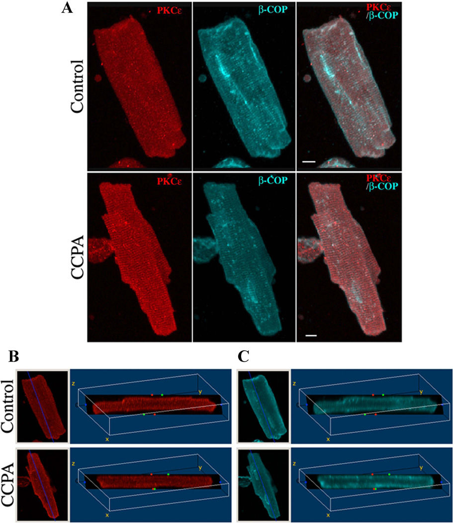
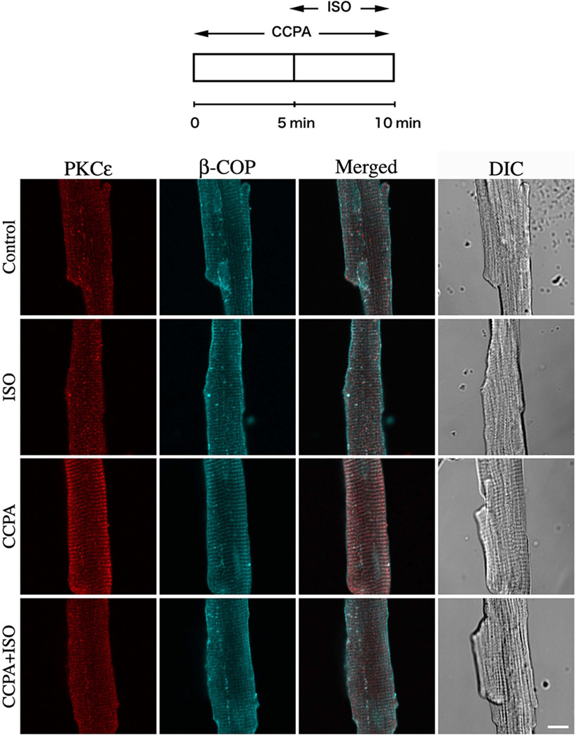
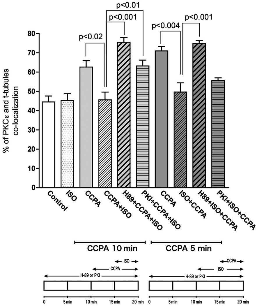
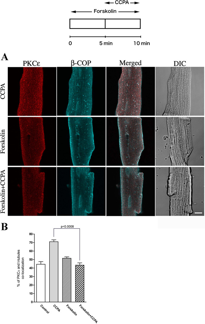
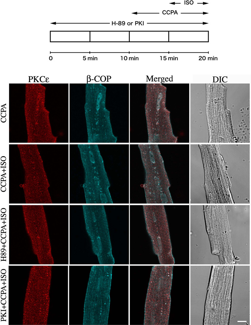
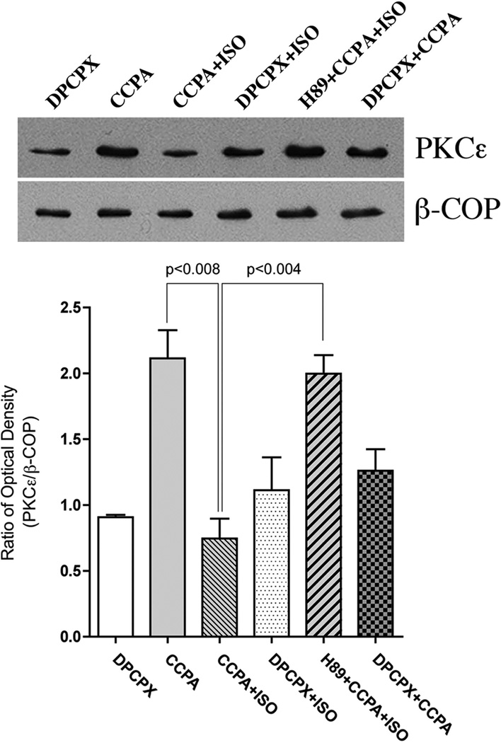
Adenosine A(1) receptor (A(1)R)-induced translocation of PKCε to transverse (t) tubular membranes in isolated rat cardiomyocytes is associated with a reduction in β(1)-adrenergic-stimulated contractile function. The PKCε-mediated activation of protein kinase D (PKD) by endothelin-1 is inhibited by β(1)-adrenergic stimulated protein kinase A (PKA) suggesting a similar mechanism of A(1)R signal transduction modulation by adrenergic agonists may exist in the heart. We have investigated the influence of β(1)-adrenergic stimulation on PKCε translocation elicited by A(1)R. Immunofluorescence imaging and Western blotting with PKCε and β-COP antibodies were used to quantify the co-localization of PKCε and t-tubular structures in isolated rat cardiomyocytes. The A(1)R agonist CCPA increased the co-localization of PKCε and t-tubules as detected by imaging. The β(1)-adrenergic receptor agonist isoproterenol (ISO) inhibited this effect of CCPA. Forskolin, a potent activator of PKA, mimicked, and H89, a pharmacological PKA inhibitor, and PKI, a membrane-permeable PKA peptide PKA inhibitor, attenuated the negative effect of ISO on the A(1)R-mediated PKCε translocation. Western blotting with isolated intact hearts revealed an increase in PKCε/β-COP co-localization induced by A(1)R. This increase was attenuated by the A(1)R antagonist DPCPX and ISO. The ISO-induced attenuation was reversed by H89. It is concluded that adrenergic stimulation inhibits A(1)R-induced PKCε translocation to the PKCε anchor site RACK2 constituent of a coatomer containing β-COP and associated with the t-tubular structures of the heart. In that this translocation has been previously associated with the antiadrenergic property of A(1)R, it is apparent that the interactive effects of adenosine and β(1)-adrenergic agonists on function are complex in the heart.


Figures

Figure 1
Figure 1. Three-dimensional reconstruction images of PKCε in isolated cardiomyocytes
Isolated rat cardiomyocytes were incubated for 10 min in the absence (Control) or presence (CCPA) of 1 µM CCPA. Cells were fixed and stained as described in “Materials and Methods”. Panel A: A series of optical sections were collected at 0.5-µm intervals from the bottom to the top of the cell and reconstructed into composite images using software from Leica Microsystems. Panels B and C: Distribution of fluorescence in the transverse section of the 3D volume images obtained from Fig 1A as defined by the dark plane. Left image of each panel shows projections of the 3D reconstructions using maximum intensity in the xy (top view) orientation. Right image shows a single slice section image that is rotated about a fixed point (x = 110, z = −70). PKCε (Red), β-COP (Blue) and Merged. Bar = 8 µm.
Figure 2
Figure 2. Effect of β1 adrenergic agonist, ISO, on the A1R-mediated translocation of PKCε in isolated cardiomyocytes
Top schematic depicts time course of agent administration. CCPA and ISO were each administered at 2 µM. The focal plane was near the middle of the myocyte viewed by confocal microscope. PKCε (Red), β-COP (Blue) and differential interference contrast (DIC) images. Bar = 10 µm.
Figure 3
Figure 3. Statistical analysis of isolated rat cardiomyocyte images indicating co-localization of PKCε with β-COP, and used as an index of PKCε binding to RACK2 (see text)
Co-localization was determined and statistics conducted as described in “Materials and Methods”. Values are means ± SE for at least 15 cardiomyocytes. Significance is indicated at the respective p value. Bottom schematics depict two protocols for exposure of cardiomyocytes.
Figure 4
Figure 4. Effect of CCPA on PKCε-induced translocation by forskolin in isolated cardiomyocytes
(A) Top schematic depicts the time course of the administration of 20 µM forskolin and 1 µM CCPA. The focal plane was near the middle of the myocyte viewed by confocal microscope. PKCε (Red), β-COP (Blue) and DIC images. Bar = 10 µm. (B) Co-localization analysis from the images similar to those presented in (A). Values are means ± SE for at least 15 cardiomyocytes. Significance is indicated at the respective p value.
Figure 5
Figure 5. Effect of the PKA inhibitor H89 and PKI peptide on the PKCε translocation effects of CCPA and ISO in isolated rat cardiomyocytes
Top schematic depicts the time course of administration to 10 µM H89 or 10 µM PKI peptide, 1 µM CCPA and 2 µM ISO. The focal plane was near the middle of the myocyte viewed by confocal microscope. PKCε (Red), β-COP (Blue) and DIC images. Bar = 10 µm.
Figure 6
Figure 6. Effect of various treatments on the translocation of PKCε to the β-COP containing membranes in the perfused rat heart as determined with immunoblotting
Membrane fractions of treated hearts were subjected to SDS-PAGE, followed by immunoblotting with PKCε Ab and β-COP Ab. Agents were administered as follows: 10 µM H89, 1 µM CCPA, 10 nM ISO and 0.1 µM 8-cyclopentyl-1,3-dipropylxanthine (DPCPX). The amount of PKCε was quantitatively determined by scanning densitometry (NIH image program). The densities of PKCε bands were normalized to those of the β-COP bands. The data are interpreted to reflect the binding of PKCε to its anchor protein β’-COP (RACK2), as described in text. The values shown are means ± SE from three independent hearts. Significance is indicated at the respective p values.

References

    1. Csukai M, Chen C-H, DeMatteis MA, Mochly-Rosen D. The coatomer protein β'COP: a selective binding protein (RACK) for epsilon protein kinase C. J Biol Chem. 1997;272:29200–29206. - PubMed
    1. Disatnik M-H, Buraggi G, Mochly-Rosen D. Localization of protein kinase C isozymes in cardiac myocytes. Exp Cell Res. 1994;210:287–297. - PubMed
    1. Dobson JG., Jr Reduction by adenosine of the isoproterenol-induced increase in cyclic adenosine 3',5'-monophosphate formation and glycogen phosphorylase activity in rat heart muscle. Circ Res. 1978;43:785–792. - PubMed
    1. Dobson JG., Jr Adenosine reduces catecholamine contractile responses in oxygenated and hypoxic atria. Am J Physiol. 1983a;245:H468–H474. - PubMed
    1. Dobson JG., Jr Mechanism of adenosine inhibition of catecholamine-induced elicited responses in heart. Circ Res. 1983b;52:151–160. - PubMed

Publication types

MeSH terms

Substances

LinkOut - more resources