

Soluble adenylyl cyclase antibody profile as a diagnostic adjunct in the assessment of pigmented lesions
- PMID: 22105816
- PMCID: PMC3387488
- DOI: 10.1001/archdermatol.2011.338
Soluble adenylyl cyclase antibody profile as a diagnostic adjunct in the assessment of pigmented lesions
Abstract
Objective: To investigate the usefulness of a novel marker for melanocytic proliferations.
Design: Using a novel monoclonal antibody against soluble adenylyl cyclase (sAC), various benign and malignant melanocytic proliferations were immunostained.
Setting: Weill Medical College of Cornell University dermatopathology laboratory.
Main outcome measures: The results were qualitative, not quantifiable.
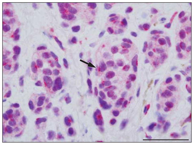
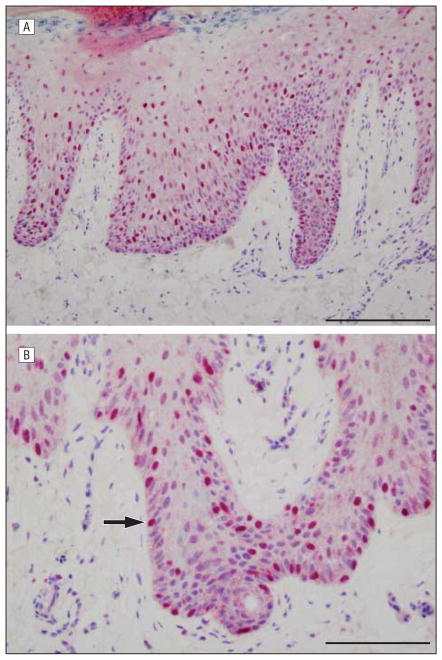
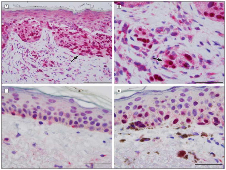
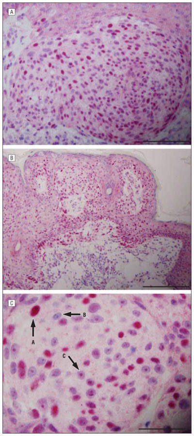
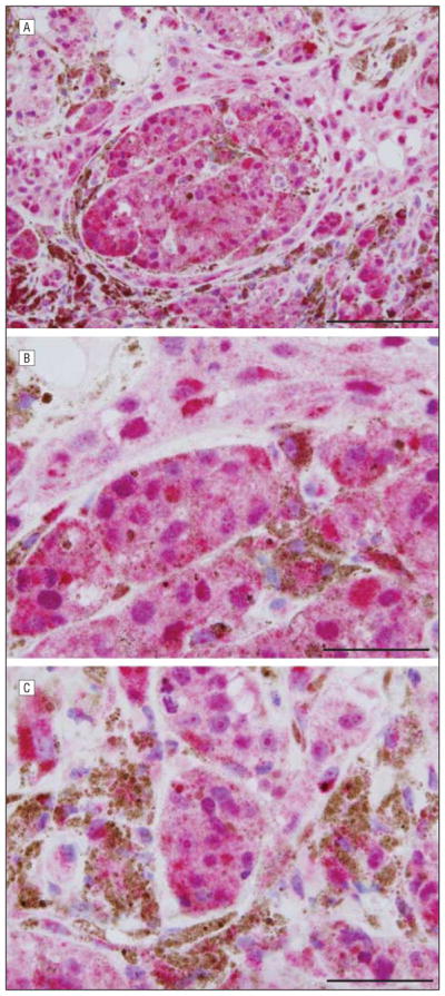
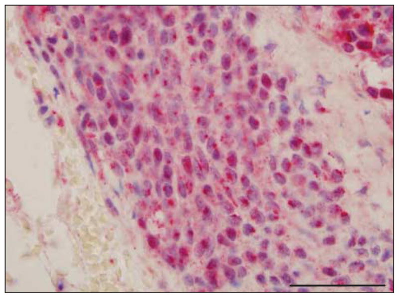
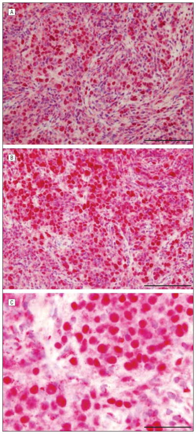
Results: The sAC immunostaining produced distinctive patterns that paralleled melanomagenesis. At one pole of the spectrum were benign nevi, including atypical nevi of special sites and recurrent nevi showing a distinct pattern of dotlike Golgi staining, while at the opposite pole was melanoma, in which many cells demonstrated an intense pannuclear expression pattern, often accompanied by loss of the Golgi expression pattern. Melanomas of lentigo maligna and acral lentiginous subtypes exhibited the most striking pannuclear expression, while nodular melanomas showed the least, although with supervening enhanced diffuse cytoplasmic expression. Loss of the Golgi expression pattern was a feature of malignant melanoma.
Conclusion: The sAC expression pattern is complex but seems discriminatory, with distinctive and variable staining patterns according to the nature of the lesion biopsied.
Figures
References
-
- Crowson AN, Magro CM, Mihm MC. The Melanocytic Proliferations: a Comprehensive Textbook of Pigmented Lesions. New York, NY: Wiley-Liss; 2001.
-
- Lebe B, Pabuççuoğlu U, Ozer E. The significance of Ki-67 proliferative index and cyclin D1 expression of dysplastic nevi in the biologic spectrum of melanocytic lesions. Appl Immunohistochem Mol Morphol. 2007;15(2):160–164. - PubMed
-
- Li LX, Crotty KA, McCarthy SW, Palmer AA, Kril JJ. A zonal comparison of MIB1-Ki67 immunoreactivity in benign and malignant melanocytic lesions. Am J Dermatopathol. 2000;22(6):489–495. - PubMed
Publication types
MeSH terms
Substances
Grants and funding
LinkOut - more resources
Full Text Sources
Other Literature Sources
Medical
