. 2012 Apr;113(4):1265-73.
doi: 10.1002/jcb.23459.

Comprehensive analysis of titin protein isoform and alternative splicing in normal and mutant rats

Affiliations

Comprehensive analysis of titin protein isoform and alternative splicing in normal and mutant rats

Shijun Li et al. J Cell Biochem. 2012 Apr.

Abstract

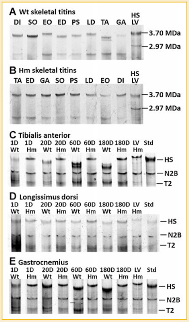
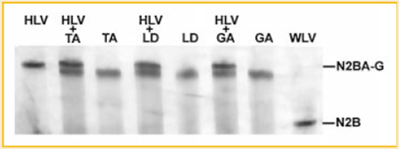
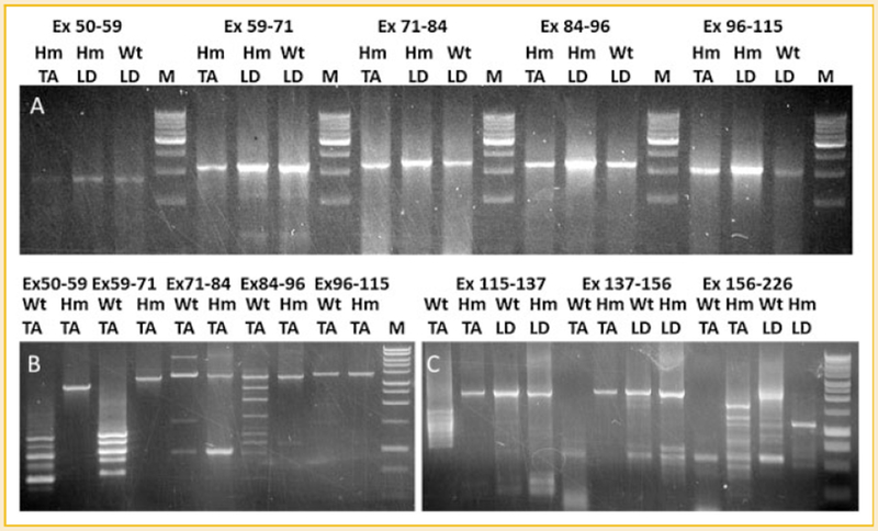
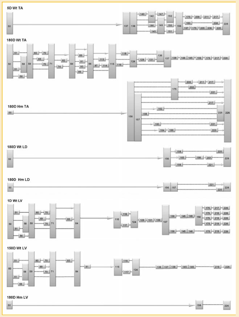
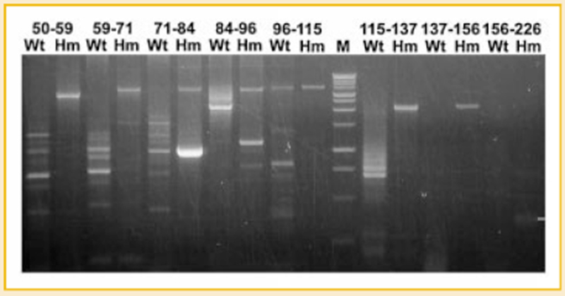
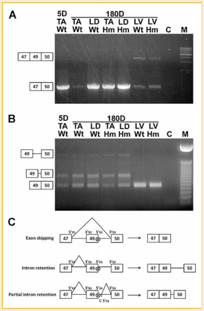
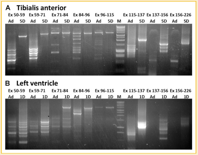
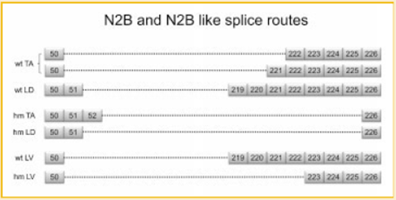
Titin is a giant protein with multiple functions in cardiac and skeletal muscles. Rat cardiac titin undergoes developmental isoform transition from the neonatal 3.7 MDa N2BA isoform to primarily the adult 2.97 MDa N2B isoform. An autosomal dominant mutation dramatically altered this transformation. Titins from eight skeletal muscles: Tibialis Anterior (TA), Longissimus Dorsi (LD) and Gastrocnemius (GA), Extensor Digitorum Longus (ED), Soleus (SO), Psoas (PS), Extensor Oblique (EO), and Diaphram (DI) were characterized in wild type and in homozygous mutant (Hm) rats with a titin splicing defect. Results showed that the developmental reduction in titin size is eliminated in the mutant rat so that the titins in all investigated skeletal muscles remain large in the adult. The alternative splicing of titin mRNA was found repressed by this mutation, a result consistent with the large titin isoform in the mutant. The developmental pattern of titin mRNA alternative splicing differs between heart and skeletal muscles. The retention of intron 49 reveals a possible mechanism for the absence of the N2B unique region in the expressed titin protein of skeletal muscle.


Figures

Fig. 1.
Fig. 1.
Titin isoforms in different skeletal muscles and the developmental transition of titin isoform in wild type and homozygote animals. A: The size of titin varies in different skeletal muscles of wild type rat; (B) The size of titin remains the same in all mutant skeletal muscles. A mixture of human soleus (HS, titin size of 3.7 MDa) and rat left ventricle (LV, titin size of 2.97 MDa) is included as a size ruler; (C–E) Titin isoforms in TA, LD, and GA was investigated at four time points: 1 day (D), 20, 60, and 180 days for both genotypes. Results show that titin in Wt TA and Wt GA changed dramatically from 1 to 180 days where as the Wt LD titin isoforms barely changed during this period. Titin size in mutant skeletal muscles remained constant during development. Wild type LV was mixed with each sample as a migration distance marker. HS, human Soleus; N2B, the major adult ventricle titin isoform; T2, titin proteolytic fragment.
Fig. 2.
Fig. 2.
Size comparison of mutant cardiac titin and mutant skeletal titin. Hm skeletal titins from TA, LD, and GA are used to compare with Hm cardiac titin, showing that Hm skeletal titin is smaller than Hm cardiac titin. HLV, Hm left ventricle; TA, (from Hm); LD, (from Hm); GA, (from Hm); WLV, wild type left ventricle.
Fig. 3.
Fig. 3.
Splicing analysis of titin mRNA in wild type and mutant skeletal muscles. cDNAs from Wt TA, Hm TA, Wt LD, and Hm LD are amplified by primer sets: 50F-59R, 59F-71R, 71F-84R, 84F-96R 96F-115R, 115F-137R, 137F-156R, and 156F-226R for the corresponding exons (Ex, exon; F, forward primer; R, reverse primer). A: only constitutive splicing can be found in the middle Ig region (exon 50-115) of Hm TA, Wt LD, and Hm LD. B: alternative splicing of middle Ig region can be found in Wt TA but only constitutive splicing in Hm TA. C: Only alternative splicing can be detected in the PEVK region (exon 115-226) of Wt TA but constitutive splicing exists in wt LD, Hm LD, and Hm TA from exon 115 to 156; the region from exon 156 to 226 is alternatively spliced in all tested tissues.
Fig. 4.
Fig. 4.
Summary of the exon profile of titin mRNA corresponding to middle Ig and PEVK region in different tissues. The colored boxes are exons (with their numbers), the solid arrows indicate consecutive exons. The dotted lines denote direct attachment between the adjacent numbered exons, the information between exon 156 to 226 in 180D Hm LV is missing.
Fig. 5.
Fig. 5.
Splicing analysis of titin mRNA in wild type and mutant cardiac muscles. Numbers correspond to the exons for the primer pairs. Alternative splicing dominates in wild type cardiac titin mRNA but only constitutive splicing was detected in the mutant. The bright band found in 71-84 Hm with size smaller than constitutively spliced product does not contain titin sequence.
Fig. 6.
Fig. 6.
The skipping of exon 49 and retention of intron 49. A: Exon 49 is absent from skeletal muscles but present in both wild type and mutant left ventricle (LV). Exons are illustrated by boxes. Exon 48 has not been found in rat titin mRNA; the constitutive splicing pattern is exon 47-49-50 in cardiac muscles and alternative splicing of exon 47 to 50 in skeletal muscles. B: Complete intron 49 retention and partial intron 49 retention occurs in titin mRNA of skeletal muscles but not in cardiac muscles. Solid lines connecting the exons indicate complete and partial intron retention (from the sequenced products). C: Schematic diagram of exon 49 skipping and intron 49 retention, 5′ splice site (ss) and 3′ ss of each exon are illustrated, “C 5′ ss” in partial intron 49 retention refers to cryptic 5′ ss.
Fig. 7.
Fig. 7.
Developmental changes of alternative splicing of titin mRNA corresponding to middle Ig and PEVK regions. A: splicing of titin mRNA in adult TA and 5 day neonatal TA. B: Splicing of titin mRNA in adult LV and 1 day neonatal LV. Results show that titin mRNA in the adult undergoes more extensive alternative splicing than the neonatal. Titin mRNA in neonatal, LV is alternatively spliced earlier than that in TA.
Fig. 8.
Fig. 8.
N2B and N2B like splice routes in various tissues. Boxes with numbers correspond to exons. The dotted lines denote direct attachment between the numbered exons.

References

    1. Bang ML, Centner T, Fornoff F, Geach AJ, Gotthardt M, McNabb M, Witt CC, Labeit D, Gregorio CC, Granzier H, Labeit S. 2001. The complete gene sequence of titin, expression of an unusual approximately 700-kDa titin isoform, and its interaction with obscurin identify a novel Z-line to I-band linking system. Circ Res 89:1065–1072. - PubMed
    1. Berget SM. 1995. Exon recognition in vertebrate splicing. J Biol Chem 270: 2411–2414. - PubMed
    1. Centner T, Yano J, Kimura E, McElhinny AS, Pelin K, Witt CC, Bang ML, Trombitas K, Granzier H, Gregorio CC, Sorimachi H, Labeit S. 2001. Identification of muscle specific ring finger proteins as potential regulators of the titin kinase domain. J Mol Biol 306:717–726. - PubMed
    1. Freiburg A, Trombitas K, Hell W, Cazorla O, Fougerousse F, Centner T, Kolmerer B, Witt C, Beckmann JS, Gregorio CC, Granzier H, Labeit S. 2000. Series of exon-skipping events in the elastic spring region of titin as the structural basis for myofibrillar elastic diversity. Circ Res 86:1114–1121. - PubMed
    1. Funatsu T, Higuchi H, Ishiwata S. 1990. Elastic filaments in skeletal muscle revealed by selective removal of thin filaments with plasma gelsolin. J Cell Biol 110:53–62. - PMC - PubMed

Publication types