Review
. 2012 Mar;212(3):243-55.
doi: 10.1530/JOE-11-0296. Epub 2011 Nov 21.

Signaling regulation of fetoplacental angiogenesis

Affiliations
Review

Signaling regulation of fetoplacental angiogenesis

Kai Wang et al. J Endocrinol. 2012 Mar.

Abstract

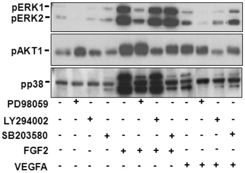
During normal pregnancy, dramatically increased placental blood flow is critical for fetal growth and survival as well as neonatal birth weights and survivability. This increased blood flow results from angiogenesis, vasodilatation, and vascular remodeling. Locally produced growth factors including fibroblast growth factor 2 (FGF2) and vascular endothelial growth factor A (VEGFA) are key regulators of placental endothelial functions including cell proliferation, migration, and vasodilatation. However, the precise signaling mechanisms underlying such regulation in fetoplacental endothelium are less well defined, specifically with regard to the interactions amongst protein kinases (PKs), protein phosphatase, and nitric oxide (NO). Recently, we and other researchers have obtained solid evidence showing that different signaling mechanisms participate in FGF2- and VEGFA-regulated fetoplacental endothelial cell proliferation and migration as well as NO production. This review will briefly summarize currently available data on signaling mediating fetoplacental angiogenesis with a specific emphasis on PKs, ERK1/2, AKT1, and p38 MAPK and protein phosphatases, PPP2 and PPP3.


Conflict of interest statement

Declaration of interest

The authors declare that there is no conflict of interest that could be perceived as prejudicing the impartiality of the research reported.

Figures

Figure 1
Figure 1
Effects of PD98059 and LY294002 on FGF2- and VEGFA-Stimulated OFPAE Cell Migration. Cell migration was measured using a Multiwell BD Falcon FluoroBlok Insert System (8.0-um pores, BD Biosciences). Cells were treated with 10 ng/ml of FGF2 or VEGFA in the absence or presence of PD98059 or LY294002 (1 hr of pretreatment). Cells were counted after 16 hr of treatment. A) Representative images are shown, in which concentrations of PD98059 and LY294002 were at 40 and 6 μM, respectively. Bar = 100 μm. B) Data are expressed as means ± SEM % of the controls. All data were analyzed using one-way ANOVA. When an F test was significant, data were compared to the control by Bonferroni’s multiple comparisons or Student’s t test. *differ from the control (n = 5; p ≤ 0.05).
Figure 2
Figure 2
Effects of SB203580 on FGF2- and VEGFA-Stimulated Cell Proliferation and FGF2-Stimulated Cell Migration in OFPAE Cells. A) A time course for FGF2- and VEGFA-induced phosphorylation of p38 MAPK. Note: 1) Both FGF2 and VEGFA (10 ng/ml) rapidly (~ 10 min) induced (p ≤ 0.05) overall p38 MAPK phosphorylation; 2) FGF2 appeared to be much more potent in inducing p38 MAPK phosphorylation as compared to VEGFA; and 3) FGF2 and VEGFA did not significantly alter protein levels of total p38 MAPK (p38). Data shown were collected after 10 min of FGF2 and VEGFA stimulation, pooled from all three p38 MAPK isoforms, and analyzed as described above. Data normalized to total p38 are expressed as means ± SEM fold of the controls. *differ from the control (n = 5; p ≤ 0.05). B) Effects of SB203580 on OFPAE cell proliferation and migration. Cell proliferation assay was carried out using a crystal violet method. Cells were treated with 10 ng/ml of FGF2 or VEGFA in the absence or presence of SB203580 (1 hr pretreatment). Cells were counted after 72 or 16 hr of treatments, respectively for cell proliferation and migration assays.
Figure 2
Figure 2
Effects of SB203580 on FGF2- and VEGFA-Stimulated Cell Proliferation and FGF2-Stimulated Cell Migration in OFPAE Cells. A) A time course for FGF2- and VEGFA-induced phosphorylation of p38 MAPK. Note: 1) Both FGF2 and VEGFA (10 ng/ml) rapidly (~ 10 min) induced (p ≤ 0.05) overall p38 MAPK phosphorylation; 2) FGF2 appeared to be much more potent in inducing p38 MAPK phosphorylation as compared to VEGFA; and 3) FGF2 and VEGFA did not significantly alter protein levels of total p38 MAPK (p38). Data shown were collected after 10 min of FGF2 and VEGFA stimulation, pooled from all three p38 MAPK isoforms, and analyzed as described above. Data normalized to total p38 are expressed as means ± SEM fold of the controls. *differ from the control (n = 5; p ≤ 0.05). B) Effects of SB203580 on OFPAE cell proliferation and migration. Cell proliferation assay was carried out using a crystal violet method. Cells were treated with 10 ng/ml of FGF2 or VEGFA in the absence or presence of SB203580 (1 hr pretreatment). Cells were counted after 72 or 16 hr of treatments, respectively for cell proliferation and migration assays.
Figure 3
Figure 3
Effects of PD98059, LY294002, and SB203580 on Phosphorylation of ERK1/2, AKT1 and p38 MAPK Induced by FGF2 and VEGFA in OFPAE Cells. After 16 hr of serum starvation, cells were treated with 10 ng/ml of FGF2 or VEGFA for 10 min in the absence or presence of PD 98059 (20 μM), LY294002 (5 μM), or SB203580 (10 μM) (1 hr pretreatment). Proteins (20μg/lane) were subjected to Western blotting using antibody against phospho-specific ERK1/2 (pERK1/2; 1:2000), AKT1 (pAKT1; 1:1000) or p38 MAPK (pp38; 1:1000).
Figure 4
Figure 4
A Proposed Model of the Signal Transduction Pathways for FGF2- and VEGFA-Stimulated Proliferation and Migration in OFPAE Cells. In this working model, we propose that FGF2 and VEGFA activate ERK1/2, AKT1, and p38 MAPK, which in turn increase eNOS protein expression and/or eNOS activity, increasing NO production. This increased NO as an intracellular signaling modulates FGF2- and VEGFA-stimulated cell proliferation and migration. Inhibition of PPP3CA, but not PPP2CA enhances VEGFA-, but not FGF2-stimulated cell proliferation, while failing to affect FGF2- and VEGFA-induced activation of ERK1/2 and AKT1, suggesting that yet to be identified signaling molecules play an important role in FGF2- and VEGFA-stimulated cell proliferation after knockdown of PPP3CA in OFPAE cells.

References

    1. Aitsebaomo J, Portbury AL, Schisler JC, Patterson C. Brothers and sisters: molecular insights into arterial-venous heterogeneity. Circulation Research. 2008;103:929–939. - PMC - PubMed
    1. Alessi DR, Gomez N, Moorhead G, Lewis T, Keyse SM, Cohen P. Inactivation of p42 MAP kinase by protein phosphatase 2A and a protein tyrosine phosphatase, but not CL100, in various cell lines. Current Biology. 1995;5:283–295. - PubMed
    1. Alexander G. Birth weight of lambs. Influences and consequences. In: Elliott K, Knight J, editors. Ciba Foundation Symposium 27: Size at Birth. New York: Elsevier Publisher; 1974.
    1. Aramburu J, Heitman J, Crabtree GR. Calcineurin: a central controller of signaling in eukaryotes. EMBO Reports. 2004;5:343–348. - PMC - PubMed
    1. Babaei S, Teichert-Kuliszewska K, Monge JC, Mohamed F, Bendeck MP, Stewart DJ. Role of nitric oxide in the angiogenic response in vitro to basic fibroblast growth factor. Circulation Research. 1998;82:1007–1015. - PubMed

Publication types