Review
. 2012 Jan 1;590(1):49-61.
doi: 10.1113/jphysiol.2011.221689. Epub 2011 Nov 21.

Redefining the classification of AMPA-selective ionotropic glutamate receptors

Affiliations
Review

Redefining the classification of AMPA-selective ionotropic glutamate receptors

Derek Bowie. J Physiol. .

Abstract

AMPA-type ionotropic glutamate receptors (iGluRs) represent the major excitatory neurotransmitter receptor in the developing and adult vertebrate CNS. They are crucial for the normal hardwiring of glutamatergic circuits but also fine tune synaptic strength by cycling into and out of synapses during periods of sustained patterned activity or altered homeostasis. AMPARs are grouped into two functionally distinct tetrameric assemblies based on the inclusion or exclusion of the GluA2 receptor subunit. GluA2-containing receptors are thought to be the most abundant AMPAR in the CNS, typified by their small unitary events, Ca(2+) impermeability and insensitivity to polyamine block. In contrast, GluA2-lacking AMPARs exhibit large unitary conductance, marked divalent permeability and nano- to micromolar polyamine affinity. Here, I review evidence for the existence of a third class of AMPAR which, though similarly Ca(2+) permeable, is characterized by its near-insensitivity to internal and external channel block by polyamines. This novel class of AMPAR is most notably found at multivesicular release synapses found in the avian auditory brainstem and mammalian retina. Curiously, these synapses lack NMDA-type iGluRs, which are conventionally associated with controlling AMPAR insertion. The lack of NMDARs suggests that a different set of rules may govern AMPAR cycling at these synapses. AMPARs with similar functional profiles are also found on some glial cells suggesting they may have a more widespread distribution in the mammalian CNS. I conclude by noting that modest changes to the ion-permeation pathway might be sufficient to retain divalent permeability whilst eliminating polyamine sensitivity. Consequently, this emerging AMPAR subclass need not be assembled from novel subunits, yet to be cloned, but could simply occur by varying the stoichiometry of existing proteins.


Figures
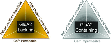
Figure 1
Figure 1. The inter-relationship between the GluA2 subunit, ion permeation and channel block
Left, GluA2-lacking AMPARs are Ca2+ permeable and strongly blocked by both external and cytoplasmic polyamines. The latter mechanism accounts for the occurrence of inward rectification. Right, in contrast, GluA2-containing AMPARs are thought to be divalent impermeable and insensitive to both external and internal polyamine block with small single channel conductance.
Figure 2
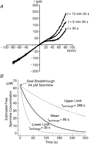
Figure 2. Washout of cytoplasmic polyamines is variable
A, a series of current–voltage relationships of AMPAR responses recorded at different time points of a whole-cell recording from a single cerebellar granule cell. Following seal breakthrough, the initial inward rectification observed at positive membrane potentials dissipated over the next 10 min reflecting washout of endogenous polyamines. Adapted from Kamboj et al. (1995) with permission. B, in HEK 293 cells expressing GluK2 kainate receptor channels, the lower and upper limits of polyamine washout were estimated using internal solutions that contained 20 mm ATP, to chelate endogenous polyamines, and solutions which lacked it, respectively. By fitting whole-cell membrane currents with a modified Woodhull model of channel block, it was possible to calculate the free concentration of spermine (ordinate axis) from the degree of rectification at different time points (abscissa) after breakthrough (see Bowie & Mayer, 1995 for details). Upper and lower estimates of polyamine exchange time constants (τexchange) were calculated to be 30 s and 288 s, respectively. Note that the mean τexchange is weighted in that 5 out of 8 recordings were obtained with internals solutions containing 20 mm ATP. Adapted from Bowie & Mayer (1995) with permission from Elsevier.
Figure 3
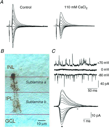
Figure 3. Not all Ca2+-permeable AMPA receptors exhibit inward rectification
A, nerve-evoked synaptic AMPAR currents elicited by chick nMAG neurons are outwardly rectifying in solutions rich in Na+ (left panel) but yet are highly permeable to external Ca2+ ions (right panel). Traces are adapted from Otis et al. (1995) with permission. B and C, likewise, AII amacrine cells (B) of the rodent retina express Ca2+-permeable AMPARs that also show no sign of inward rectification at positive membrane potentials. Photomicrograph in B is adapted from Osswald et al. (2007) and traces in C are adapted from Veruki et al. (2003) with permission.
Figure 4
Figure 4. Eternal polyamine block can be uncoupled from AMPA receptor divalent ion permeability
A, typical membrane currents elicited by synaptic AMPARs expressed by AII amacrine cells after eye-opening are resistant to supramaximal concentrations of the polyamine toxin, PhTX (right panel). B, likewise, Co2+ staining elicited by AMPA receptor stimulation reveals that the divalent permeability of these receptors is not blocked by a range of known channel blockers. Data adapted from Osswald et al. (2007) with permission.
Figure 5
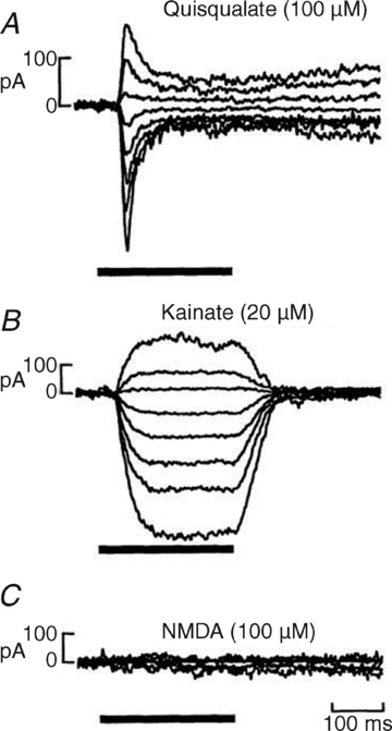
Figure 5. Cells expressing polyamine-insensitive, Ca2+-permeable AMPA receptors often lack NMDA receptors
Whole-cell membrane currents recorded from salamander retinal bipolar cells following a 250 ms application of the AMPAR agonists, quisqualate and kainate (A and B) and the NMDA receptor agonist, NMDA (C) at a range of membrane potentials (−95 mV to +45 mV). Note that although bipolar cells respond robustly to the AMPAR agonists there is no effect of NMDA. Data adapted from Gilbertson et al. (1991) and reprinted with permission from AAAS.

References

    1. Aizenman CD, Munoz-Elias G, Cline HT. Visually driven modulation of glutamatergic synaptic transmission is mediated by the regulation of intracellular polyamines. Neuron. 2002;34:623–634. - PubMed
    1. Almasieh A, Wilson A, Morquette B, Vargas J, Di PA. The molecular basis of retinal ganglion cell death in glaucoma. Prog Retin Eye Res. 2011 (in Press) - PubMed
    1. Bahring R, Bowie D, Benveniste M, Mayer ML. Permeation and block of rat GluR6 glutamate receptor channels by internal and external polyamines. J Physiol. 1997;502:575–589. - PMC - PubMed
    1. Banke TG, Bowie D, Lee H, Huganir RL, Schousboe A, Traynelis SF. Control of GluR1 AMPA receptor function by cAMP-dependent protein kinase. J Neurosci. 2000;20:89–102. - PMC - PubMed
    1. Beattie EC, Stellwagen D, Morishita W, Bresnahan JC, Ha BK, Von ZM, Beattie MS, Malenka RC. Control of synaptic strength by glial TNFa. Science. 2002;295:2282–2285. - PubMed

Publication types

MeSH terms

Substances

LinkOut - more resources