

Review
doi: 10.1113/jphysiol.2011.213850.
Epub 2011 Nov 21.
Structure and function of glutamate receptor amino terminal domains
Affiliations
- PMID: 22106178
- PMCID: PMC3300046
- DOI: 10.1113/jphysiol.2011.213850
Item in Clipboard
Review
Structure and function of glutamate receptor amino terminal domains
J Physiol.
.
Abstract
The amino terminal domain (ATD) of ionotropic glutamate receptor (iGluR) subunits resides at the extracellular region distal to the membrane. The ATD is structurally and functionally the most divergent region of the iGluR subunits. Structural studies on full-length GluA2 and the ATDs from three iGluR subfamilies have shed light on how the ATD facilitates subunit assembly, accommodates allosteric modulator compounds, and controls gating properties. Here recent developments in structural and functional studies on iGluR ATDs are reviewed.
Figures
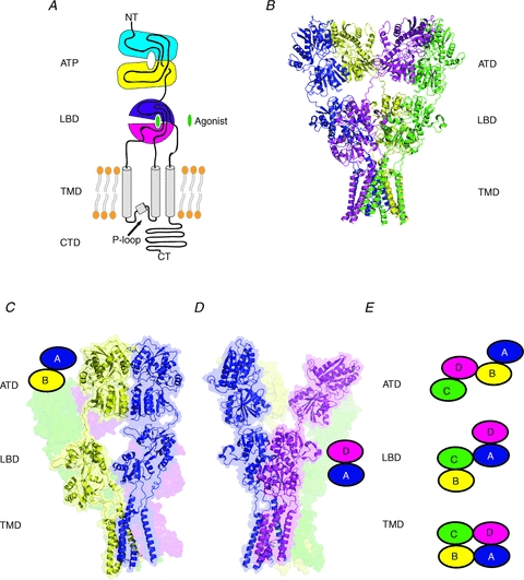
A, iGluR subunits are composed of distinct domains including the amino terminal domain (ATD), ligand-binding domain (LBD), transmembrane domain (TMD), and carboxyl terminal domain (CTD). B, crystal structure of the homotetrameric full-length GluA2 receptors (PDB code: 3KG2) showing the pattern of subunit arrangement and domain organization in the tetrameric assembly (Sobolevsky et al. 2009). The four subunits (A–D) are coloured as blue (A), yellow (B), green (C) and magenta (D). C–E, ATD dimers (panel C) and LBD dimers (panel D) are formed by an A–B (shown as a cartoon) or C–D pair and A–D (shown as a cartoon) or B–C pair, respectively. This results in a crossover of the dimer pairs in the ATD and LBD sections. Panels C, D, and E are modified from Hansen et al. (2010) with permission from the American Society for Pharmacology and Experimental Therapeutics.
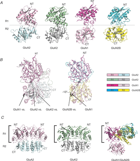
A, structures of ATD monomers from the AMPA, kainate and NMDA receptor subfamilies. The overall architecture of iGluR ATDs is shaped like a bi-lobed clamshell composed of the upper lobe (R1) and the lower lobe (R2), coloured differently. The structures (PDB codes are 3H5V, 3H6G, 3QEK and 3JPYB for GluA2, GluK2, GluN1 and GluN2B, respectively) are aligned with the similar R1 orientation. B, distinct R1–R2 orientation in NMDA receptors. Superposition of the R1 domains shows that the ATD clamshells from NMDA receptor subunits (both GluN1 and GluN2B) are substantially ‘twisted’ compared to those from non-NMDA receptors. The pivotal points for the R1–R2 twist are displayed as grey spheres. C, comparison of GluA2 ATD homodimer, GluK2 ATD homodimer, and GluN1–GluN2B ATD heterodimer. The R1s of ATDs on the left (square bracket; GluN1 R1 in GluN1–GluN2B ATD heterodimer) are similarly oriented. Note a substantial difference in the subunit orientation of GluN1–GluN2B ATDs compared to those of GluA2 or GluK2 ATDs. Panels B and C are modified from Karakas et al. (2011).
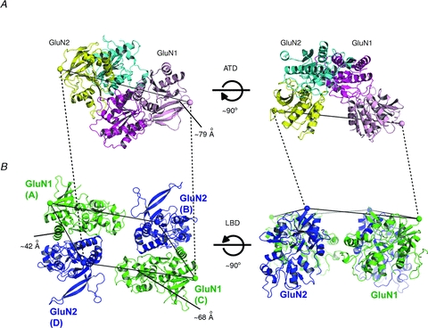
A, GluN1–GluN2B ATD dimer viewed from the C-terminal ends (spheres; left panel) or the side of the C-termini. The colour code of GluN1 and GluN2B is as in Fig. 2. B, tetrameric model of NMDA receptor LBDs assuming the GluN1–GluN2–GluN1–GluN2 orientation viewed from the sides of the N-terminal ends (spheres; left panel) and the side of N-termini. This model is built by superposing the top portion (domain 1) of the GluN1 and GluN2A LBD bi-lobed structures (PDB code: 2A5T) (Furukawa et al. 2005) onto the equivalent portion of the full length GluA2 receptor structure. Specifically, two GluN1 LBDs are superposed to the A and C subunits of GluA2 AMPA receptor structure in Fig. 1 whereas two GluN2 LBDs are superposed to the B and D subunits. The distance between the C-terminal ends of GluN1 and GluN2B ATDs is ∼79 Å whereas the GluN1–GluN2 distance between the N-termini of non-dimer forming LBDs is ∼68 Å.
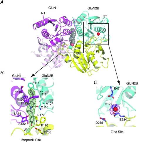
A, GluN1–GluN2B ATDs with the binding site for ifenprodil (grey sphere) at the subunit interface and for zinc (red sphere) within the GluN2B cleft. The cartoon represents the composite structure of GluN1–GluN2B ATDs in complex with ifenprodil (PDB code: 3QEL) and GluN2B ATD in complex with zinc (PDB code: 3JPY). Cyan sphere represents a water molecule at the ifenprodil binding site. B, blow-up view of the ifenprodil binding site. Binding of ifenprodil involves residues from both GluN1 and GluN2B subunits, which form hydrophobic and polar interactions. Shown in mesh is the Fo-Fc omit electron density map contoured at 3σ. A cyan sphere represents a water molecule. C, zinc binding site at the clamshell cleft of GluN2B ATD. His127 and Glu284 coordinate directly to zinc. Glu47 and Asp265 are proximal to the zinc binding site and have been previously shown to affect zinc sensitivity (Rachline et al. 2005). Water molecules that are likely to be present but not visible in this crystal structure due to limited resolution of the crystallographic data may play an important role in zinc coordination along with Glu47 and Asp265. Shown in magenta mesh is the anomalous difference Fourier map at 6σ.
References
-
- Armstrong N, Gouaux E. Mechanisms for activation and antagonism of an AMPA-sensitive glutamate receptor: crystal structures of the GluR2 ligand binding core. Neuron. 2000;28:165–181. - PubMed
-
- Armstrong N, Jasti J, Beich-Frandsen M, Gouaux E. Measurement of conformational changes accompanying desensitization in an ionotropic glutamate receptor. Cell. 2006;127:85–97. - PubMed
-
- Armstrong N, Sun Y, Chen GQ, Gouaux E. Structure of a glutamate-receptor ligand-binding core in complex with kainate. Nature. 1998;395:913–917. - PubMed
-
- Atlason PT, Garside ML, Meddows E, Whiting P, McIlhinney RA. N-Methyl-D-aspartate (NMDA) receptor subunit NR1 forms the substrate for oligomeric assembly of the NMDA receptor. J Biol Chem. 2007;282:25299–25307. - PubMed
-
- Carron C, Jullien A, Bucher B. Synthesis and pharmacological properties of a series of 2-piperidino alkanol derivatives. Arzneimittelforschung. 1971;21:1992–1998. - PubMed
Publication types
MeSH terms
Substances
Grants and funding
LinkOut - more resources
Full Text Sources
Research Materials