Review
. 2012 Jan 1;590(1):73-91.
doi: 10.1113/jphysiol.2011.223750. Epub 2011 Nov 21.

Modes of glutamate receptor gating

Affiliations
Review

Modes of glutamate receptor gating

Gabriela K Popescu. J Physiol. .

Abstract

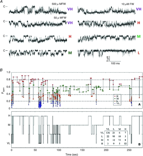
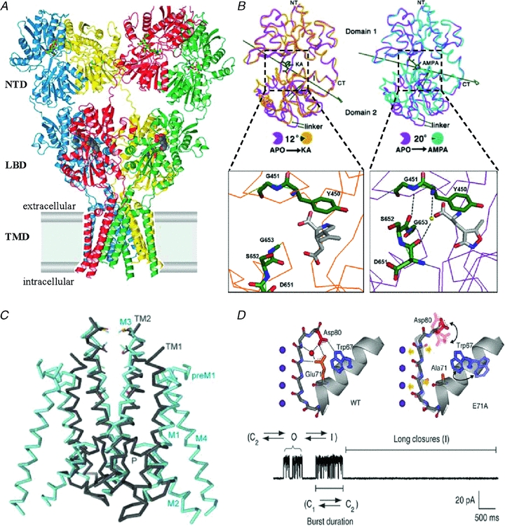
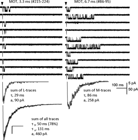
The time course of excitatory synaptic currents, the major means of fast communication between neurons of the central nervous system, is encoded in the dynamic behaviour of post-synaptic glutamate-activated channels. First-pass attempts to explain the glutamate-elicited currents with mathematical models produced reaction mechanisms that included only the most basic functionally defined states: resting vs. liganded, closed vs. open, responsive vs. desensitized. In contrast, single-molecule observations afforded by the patch-clamp technique revealed an unanticipated kinetic multiplicity of transitions: from microseconds-lasting flickers to minutes-long modes. How these kinetically defined events impact the shape of the synaptic response, how they relate to rearrangements in receptor structure, and whether and how they are physiologically controlled represent currently active research directions. Modal gating, which refers to the slowest, least frequently observed ion-channel transitions, has been demonstrated for representatives of all ion channel families. However, reaction schemes have been largely confined to the short- and medium-range time scales. For glutamate receptors as well, modal gating has only recently come under rigorous scrutiny. This article reviews the evidence for modal gating of glutamate receptors and the still developing hypotheses about the mechanism(s) by which modal shifts occur and the ways in which they may impact the time course of synaptic transmission.


Figures

Figure 1
Figure 1. Single-channel current recordings reveal modal gating
A, glutamate-activated chloride-channel activity recorded from denervated locust muscle (extracellular patch clamp). From Patlak et al. (1979); reprinted by permission from Macmillan Publishers Ltd: Nature, ©1979. B, voltage-activated calcium-channel sweeps recorded from dissociated guinea pig ventricle cells (cell attached voltage-clamp). From Hess et al. (1984); reprinted by permission from Macmillan Publishers Ltd: Nature, ©1984. C, voltage-activated sodium-channel current sweeps from Rana sartorius muscle fragments (from Patlak & Ortiz 1986). D, acetylcholine-activated channels in cultured Xenopus myocytes (cell-attached voltage-clamp) (from Auerbach & Lingle, 1986). Arrows mark sudden changes in channel kinetics that occur during a cluster of openings.
Figure 2
Figure 2. Neuronal glutamate receptor-channels have several gating modes
A, glutamate-elicited currents in dissociated rat hippocampal neurons (>2 weeks in culture; outside-out patch-clamp). From Jahr & Stevens, (1987); reprinted by permission from Macmillan Publishers Ltd: Nature, ©1987. The large conductance (50 pS) produced bursts with short (∼2 ms) or long (∼13 ms) openings indicative of two modes of gating. B, glutamate-elicited currents in cultured rat hippocampal cells (outside-out patch-clamp). Left, traces recorded at several applied glutamate concentrations, as labelled; the lowest trace shows an example of ‘high Po’ periods that occurred at all concentrations; these were excluded from kinetic analyses. Right, stability plot for mean closed time/ms (top), mean open time/ms (middle) and open probability (bottom) for an entire record (957 intervals) obtained with 20 nm glutamate (from Gibb & Colquhoun, 1991 with permission of The Royal Society).
Figure 3
Figure 3. NMDA receptors have three gating modes
A, four consecutive (non-contiguous) single-channel current traces (10 s each, GluN1/GluN2A receptors, cell-attached) illustrate shifts in opening pattern (arrows); openings are downward. B, segments (1 s each) of active time (desensitization gaps removed) sorted into three categories mirror the four switches in current pattern illustrated in A. Numbers indicate the mean for each population in the patch illustrated. C, predicted macroscopic responses for channels in each mode, elicited by a 1 ms pulse of 1 mm glutamate. For channels in each mode, peak Po is about half the equilibrium Po. Numbers indicate time constants of single exponential fits to the decay phase of the currents. Red, high (H); blue, medium (M); green, low (L); black symbols, excluded. Adapted from Popescu (2005) with permission from Springer.
Figure 4
Figure 4. High mode of gating in AMPA receptors
Currents from one GluA2(Q) channel (outside-out patches) were elicited by 200 ms applications of glutamate. Desensitization and polyamine block were reduced with CTZ (30 μm external) and ATP (internal), respectively. A, traces obtained in 60 nm glutamate illustrate openings to the four open levels (O1–4). Openings to the highest conductance level O4 were substantially longer (note different time scale); occurred at higher frequencies (bar graph) than predicted by binomial distribution; and were similar in appearance with the openings produced by 5 mm glutamate. B, left, histogram of all open durations in one patch (open bins) illustrates two exponential components (inset: time constants and areas); the fast component overlaps with the distribution of openings to the lowest conductance level, O1 (filled bins). Right, summary of time constants and areas of the fast and slow open components obtained from five patches. From Prieto & Wollmuth (2010).
Figure 5
Figure 5. Five gating modes in GluA3 receptors
Currents from one GluA3(G) channel (on-cell patches) were recorded with the indicated agonists, and 100 μm CTZ in the pipette. Regardless of agonist and its concentration, channels opened to three conductance levels (downward). During active periods (longest closed intervals removed), using a Po criterion segments fell into five categories: VH, H, M, L and VL. A, examples of single channel traces in VH, H, M and L modes. B, for one patch obtained with 50 μm FW, segments are represented in the order in which they occurred in the record and are plotted as a horizontal line according to segment Po (upper panel) or assigned gating mode (lower panel). Inset, transition matrix between modes shows that consecutive segments are more likely to have the same mode of gating than to switch mode, an indication that modal transitions occur on a much slower time scale than transitions associated with channel activation. From Poon et al. (2010), reprinted with permission from Elsevier/the Biophysical Society.
Figure 6
Figure 6. Structural hypotheses for iGluR modes
A, structure of a homo-tetrameric AMPA-type receptor in complex with antagonist (CNQX) molecules. Each subunit (blue, red, green and yellow) forms two extracellular globular domains (NTD and LBD) and a transmembrane domain (TMD). From Sobolevsky et al. (2009); reprinted by permission from Macmillan Publishers Ltd: Nature, ©2009. B, isolated GluA2 LBDs organize as two hinged lobes that can adopt slightly different positions relative to one another. Relative to the apo form (magenta), in the KA-bound form (orange) domain 2 is closer to domain 1 by 12 deg whereas in the AMPA-bound form (purple), domain 2 is closer to domain 1 by 20 deg (right panel). Also, in the AMPA-bound form two additional H-bonds form across the cleft between domains 1 and 2 due to an alternative conformation of the protein backbone around the tripeptide G653-S652-D651 (adapted from Armstrong & Gouaux, 2000 with permission from Elsevier). C, Two subunits of the KcsA channel (grey) superimposed with the membrane domain (M1–M4) of two GluA2 subunits; in the GluA2 structure the selectivity filter appears disordered (from Sobolevsky et al. 2009). D, atomic arrangements in the selectivity filter of KcsA. In the wild-type channel (left panel) a network of hydrogen bonds stabilizes the filter in a conformation that can quickly fluctuate between closed (C2) and open (O) conformations, with occasional sojourns in the C-type inactivated state (I); in the E71A KcsA mutant (right panel), increased conformational dynamics of Asp80 prevents its interaction with Trp67 and by averting filter collapse stabilizes the open state. The single channel trace illustrates fluctuations between actively gating and inactivated modes. From Cordero-Morales et al. 2006a; reprinted by permission from Macmillan Publishers Ltd: Nature Structural and Molecular Biology, ©2006.
Figure 7
Figure 7. Modal gating controls NMDA receptor macroscopic time course
About 1000 consecutive sweeps were recorded from one channel (GluN1/GluN2A receptor, outside-out patch) following brief pulses (arrow, 1 ms) of 1 mm glutamate applied at 0.2 Hz, in the presence of glycine (0.1 mm). Sweeps with similar kinetics (MOT and burst length) clustered together. Upper panels illustrate two runs of 10 consecutive sweeps, recorded within the same one-channel patch at ∼10 min interval (sweep number and MOT are indicated for each run). Below each run, is the sum trace for L- and M-sweeps, and at bottom the sum trace for all sweeps. Grey lines superimposed over summed current traces represent fits to declining exponential functions. The characteristic bi-exponential decay of an NMDA receptor macroscopic response reflects the merging of two distinct behaviours each declining with mono-exponential time course and interconverting on a min time scale. From Zhang et al. (2008).

Similar articles

Cited by

References

    1. Albright TD, Jessell TM, Kandel ER, Posner MI. Neural science: a century of progress and the mysteries that remain. Neuron. 2000;25:S1–S55. - PubMed
    1. Amico-Ruvio S, Popescu G. Stationary gating of GluN1/GluN2B receptors in intact membrane patches. Biophys J. 2010;98:1160–1169. - PMC - PubMed
    1. Amico-Ruvio SA, Murthy SE, Smith TP, Popescu GK. Zinc Effects on NMDA receptor gating kinetics. Biophys J. 2011;100:1910–1918. - PMC - PubMed
    1. Anson LC, Chen PE, Wyllie DJA, Colquhoun D, Schoepfer R. Identification of amino acid residues of the NR2A subunit that control glutamate potency in recombinant NR1/NR2A NMDA receptors. J Neurosci. 1998;18:581–589. - PMC - PubMed
    1. Anson LC, Schoepfer R, Colquhoun D, Wyllie DJ. Single-channel analysis of an NMDA receptor possessing a mutation in the region of the glutamate binding site. J Physiol. 2000;527:225–237. - PMC - PubMed

Publication types

Substances

LinkOut - more resources