

Fitness costs limit influenza A virus hemagglutinin glycosylation as an immune evasion strategy
- PMID: 22106257
- PMCID: PMC3251056
- DOI: 10.1073/pnas.1108754108
Fitness costs limit influenza A virus hemagglutinin glycosylation as an immune evasion strategy
Abstract
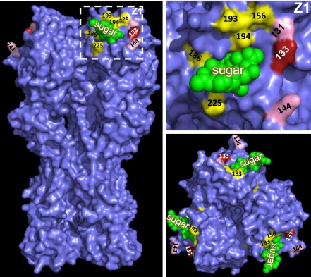
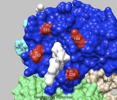
Here, we address the question of why the influenza A virus hemagglutinin (HA) does not escape immunity by hyperglycosylation. Uniquely among dozens of monoclonal antibodies specific for A/Puerto Rico/8/34, escape from H28-A2 neutralization requires substitutions introducing N-linked glycosylation at residue 131 or 144 in the globular domain. This escape decreases viral binding to cellular receptors, which must be compensated for by additional substitutions in HA or neuraminidase that enable viral replication. Sequence analysis of circulating H1 influenza viruses confirms the in vivo relevance of our findings: natural occurrence of glycosylation at residue 131 is always accompanied by a compensatory mutation known to increase HA receptor avidity. In vaccinated mice challenged with WT vs. H28-A2 escape mutants, the selective advantage conferred by glycan-mediated global reduction in antigenicity is trumped by the costs of diminished receptor avidity. These findings show that, although N-linked glycosylation can broadly diminish HA antigenicity, fitness costs restrict its deployment in immune evasion.
Conflict of interest statement
The authors declare no conflict of interest.
Figures
References
-
- Wiley DC, Skehel JJ. The structure and function of the hemagglutinin membrane glycoprotein of influenza virus. Annu Rev Biochem. 1987;56:365–394. - PubMed
-
- Dimmock NJ. Update on the neutralization of animal viruses. Rev Med Virol. 1995;5:165–179.
-
- Caton AJ, Brownlee GG, Yewdell JW, Gerhard W. The antigenic structure of the influenza virus A/PR/8/34 hemagglutinin (H1 subtype) Cell. 1982;31:417–427. - PubMed
-
- Yewdell JW, Gerhard W. Antigenic characterization of viruses by monoclonal antibodies. Annu Rev Microbiol. 1981;35:185–206. - PubMed
-
- Yewdell JW, Webster RG, Gerhard WU. Antigenic variation in three distinct determinants of an influenza type A haemagglutinin molecule. Nature. 1979;279:246–248. - PubMed
Publication types
MeSH terms
Substances
Grants and funding
LinkOut - more resources
Full Text Sources
Other Literature Sources
Medical
Molecular Biology Databases
