. 2012 Jan 12;119(2):e1-8.
doi: 10.1182/blood-2011-08-370734. Epub 2011 Nov 21.

Plasmodium falciparum STEVOR proteins impact erythrocyte mechanical properties

Affiliations

Plasmodium falciparum STEVOR proteins impact erythrocyte mechanical properties

Sohini Sanyal et al. Blood. .

Abstract

Infection of erythrocytes with the human malaria parasite, Plasmodium falciparum, results in dramatic changes to the host cell structure and morphology. The predicted functional localization of the STEVOR proteins at the erythrocyte surface suggests that they may be involved in parasite-induced modifications of the erythrocyte membrane during parasite development. To address the biologic function of STEVOR proteins, we subjected a panel of stevor transgenic parasites and wild-type clonal lines exhibiting different expression levels for stevor genes to functional assays exploring parasite-induced modifications of the erythrocyte membrane. Using this approach, we show that stevor expression impacts deformability of the erythrocyte membrane. This process may facilitate parasite sequestration in deep tissue vasculature.


Figures

Figure 1
Figure 1
Endogenous levels of stevor expression affect erythrocyte deformability. (A) Retention rates in microsphere matrices at 34 hpi for erythrocytes infected with the NF54 clonal lines B3, H4, B3B1, E10, and A12. Gray represents clones exhibiting significant expression of a least one stevor gene; and red, stevor nonexpressing clones. Measurements were performed on 2% hematocrit cultures containing 2% to 10% tightly synchronized parasites. (B) Average retention rate in microsphere matrices at 34 hpi for erythrocytes infected with clones that do not express stevor genes (red) is significantly different from that of clones expressing at least one stevor gene (gray), as measured by a Wilcoxon rank-sum test. (C) Kinetics of retention in microsphere matrices for B3 (gray) and B3B1 (red) parasite lines during parasite life cycle. Measurements were performed at 10, 16, 22, 28, 34, and 40 hpi on 2% hematocrit cultures containing 2% to 10% tightly synchronized parasites.
Figure 2
Figure 2
STEVOR proteins are involved in P falciparum–infected erythrocyte rigidity. (A) Kinetics of retention in microsphere matrices for B3 (gray), SFM+ (green), SB3 (red), and CBM-BSD (blue) parasite cultures during the parasite life cycle. Measurements were performed at 10, 16, 22, 28, 34, and 40 hpi on 2% hematocrit cultures containing 2% to 10% tightly synchronized parasites. (B) Retention rate in microsphere matrices at 16 hpi of the entire SFM+ parasite-infected erythrocyte population compared with that of the c-myc–positive SFM+ parasites. The proportion of c-myc–positive parasites before and after microsphere filtration was determined by immunofluorescence using anti–c-myc mAb and Hoechst 33342 for nuclei staining.
Figure 3
Figure 3
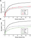
Ektacytometry analysis of erythrocyte elongation index in parasite lines. LORCA measurements of uninfected erythrocytes (black) compared with erythrocytes infected with wild-type B3 (gray) or SB3 (red) parasite lines at 34 hpi (A), and to erythrocytes infected with B3 or SFM+ (green) parasite lines at 28 hpi (B). Erythrocyte deformability is expressed as elongation index and is determined at increasing shear stress, measured in Pascal (Pa). Parasites were synchronized by MACS and concentrated to 50% parasitemia to increase sensitivity of the measurement. Error bars represent SD.
Figure 4
Figure 4
Loss of retention after shedding of episome in the SFM+ line. (A) Immunofluorescence analysis of the SFM+ parasite line cultured during 21 generations in the presence (SFM+, top panel) or absence (SFM, bottom panel) of 40nM pyrimethamine. Infected erythrocytes were stained with anti–c-myc mAb followed by anti–rat Alexa-488–conjugated IgG, and parasite nuclei were counterstained with Hoechst 33342. Pictures were taken under identical exposure conditions. Absence of c-myc expression in SFM line indicates a loss of episomal expression of the epitope-tagged STEVOR protein. (B) Scanning electron micrograph of B3, SFM+, and SFM parasite-infected erythrocytes. Right and middle panels: SFM and SFM+ infected erythrocytes with normal knobs compared with erythrocyte infected with the KAHRP-deficient B3 parasite line (left panel) in which knobs are absent. The bars represent 2 μm. (C) Kinetics of retention in microsphere matrices for erythrocytes infected with B3 (gray line), SFM+ (green continuous line), and SFM (green dotted line) during the parasite life cycle. Measurements were performed at 10, 16, 22, 28, 34, and 40 hpi on 2% hematocrit cultures containing 2% to 10% tightly synchronized parasites.
Figure 5
Figure 5
Switching of stevor expression in B3B1 results in increased erythrocyte rigidity. (A,C) Retention rate of erythrocytes infected with B3 (gray bar) and B3B1 before (B3B1 OFF, red bar) or after (B3B1 ON, green bar) switching in stevor gene family expression. Measurements were performed on 2% hematocrit cultures containing 2% to 10% tightly synchronized parasites at 34 hpi. (B,D) LORCA rigidity measurements of erythrocytes infected with B3 (gray bar) and B3B1 before (B3B1 OFF, red bar) or after (B3B1 ON, green bar) switching in stevor gene family expression. Measurements were performed on MACS-concentrated cultures containing 50% synchronized parasites at 34 hpi. (E) Analysis of transcriptional levels for the stevor gene family in B3B1 line before (B3B1 OFF, red bars) and after (B3B1 ON, green bars) switching in expression. Transcriptional analysis was performed at 26 to 28 hpi during the same life cycle as the LORCA and microsphere matrix measurements. Transcription levels were normalized to the transcription level of the seryltRNA synthetase housekeeping gene (PF07_0073).

References

    1. Maier AG, Cooke BM, Cowman AF, Tilley L. Malaria parasite proteins that remodel the host erythrocyte. Nat Rev Microbiol. 2009;7(5):341–354. - PubMed
    1. Mohandas N, Gallagher PG. Red cell membrane: past, present, and future. Blood. 2008;112(10):3939–3948. - PMC - PubMed
    1. Nash GB, O'Brien E, Gordon-Smith EC, Dormandy JA. Abnormalities in the mechanical properties of red blood cells caused by Plasmodium falciparum. Blood. 1989;74(2):855–861. - PubMed
    1. Glenister FK, Coppel RL, Cowman AF, Mohandas N, Cooke BM. Contribution of parasite proteins to altered mechanical properties of malaria-infected red blood cells. Blood. 2002;99(3):1060–1063. - PubMed
    1. Lavazec C, Sanyal S, Templeton TJ. Expression switching in the stevor and Pfmc-2TM superfamilies in Plasmodium falciparum. Mol Microbiol. 2007;64(6):1621–1634. - PubMed

Publication types

MeSH terms

Substances