. 2012 Jan;32(2):541-57.
doi: 10.1128/MCB.06032-11. Epub 2011 Nov 21.

Cdc7-Dbf4 is a gene-specific regulator of meiotic transcription in yeast

Affiliations

Cdc7-Dbf4 is a gene-specific regulator of meiotic transcription in yeast

Hsiao-Chi Lo et al. Mol Cell Biol. 2012 Jan.

Abstract

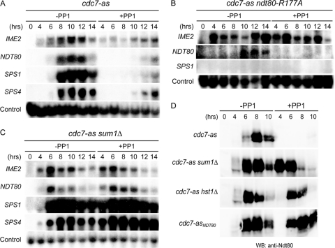
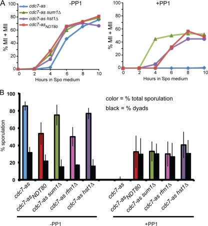
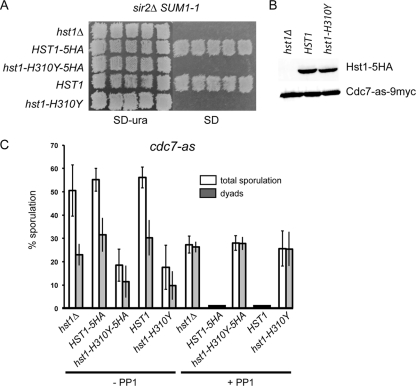
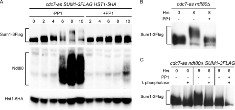
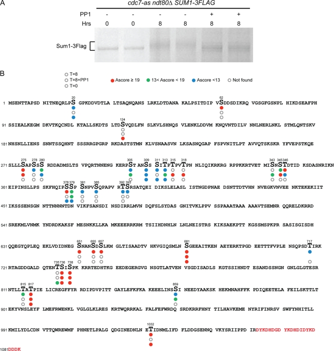
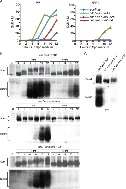
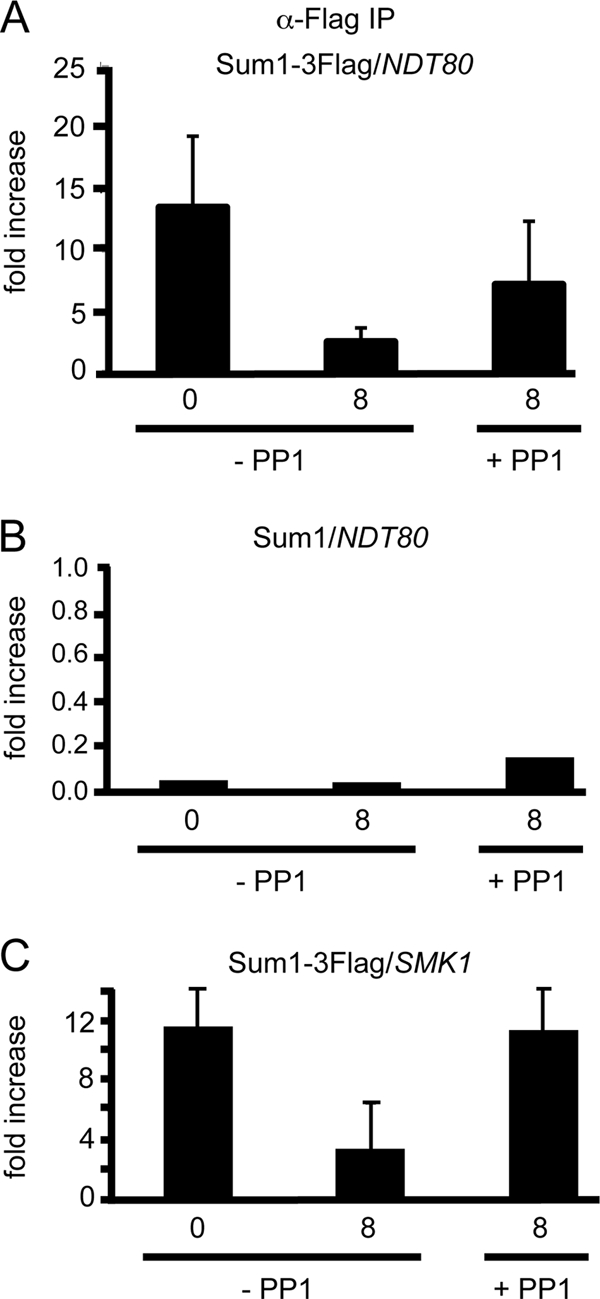
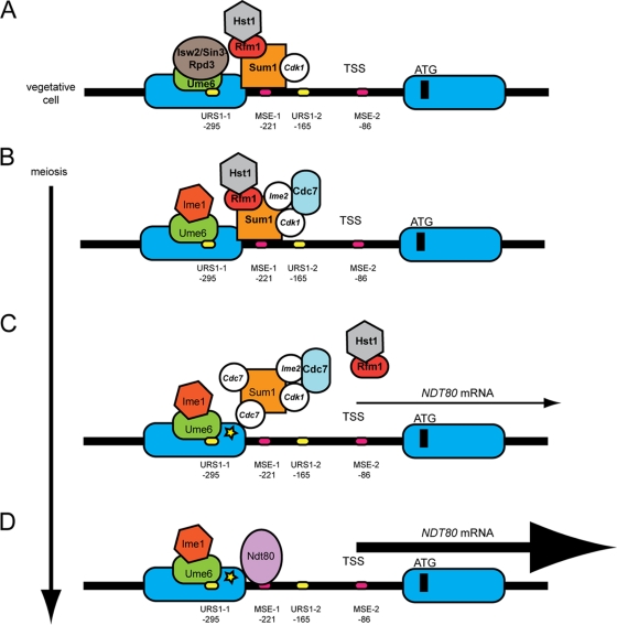
Meiosis divides the chromosome number of the cell in half by having two rounds of chromosome segregation follow a single round of chromosome duplication. The first meiotic division is unique in that homologous pairs of sister chromatids segregate to opposite poles. Recent work in budding and fission yeast has shown that the cell cycle kinase, Cdc7-Dbf4, is required for many meiosis-specific chromosomal functions necessary for proper disjunction at meiosis I. This work reveals another role for Cdc7 in meiosis as a gene-specific regulator of the global transcription factor, Ndt80, which is required for exit from pachytene and entry into the meiotic divisions in budding yeast. Cdc7-Dbf4 promotes NDT80 transcription by relieving repression mediated by a complex of Sum1, Rfm1, and a histone deacetylase, Hst1. Sum1 exhibits meiosis-specific Cdc7-dependent phosphorylation, and mass spectrometry analysis reveals a dynamic and complex pattern of phosphorylation events, including four constitutive cyclin-dependent kinase (Cdk1) sites and 11 meiosis-specific Cdc7-Dbf4-dependent sites. Analysis of various phosphorylation site mutants suggests that Cdc7 functions with both Cdk1 and the meiosis-specific kinase Ime2 to control this critical transition point during meiosis.


Figures

Fig 1
Fig 1
Early and middle meiotic gene expression in various cdc7-as diploids in the absence or presence of PP1. Diploids were transferred to Spo medium in the absence or presence of 15 μM PP1, and cells from the indicated time points were analyzed by Northern blot assays (A to C) or immunoblot assays (D). (A) cdc7-as (NH452F). (B) cdc7-as ndt80-R177A (NH932::pHL8-R177A). (C) cdc7-as sum1Δ (NH788). (D) Total cell extracts were prepared from the cdc7-as, cdc7-as sum1Δ, cdc7-as hst1Δ (NH1061), and cdc7-asNDT80 (NH452F::CUP1-NDT80) diploids at the indicated time points and probed with anti-Ndt80 antibodies.
Fig 2
Fig 2
Meiotic progression and ascus formation in various cdc7-as diploids in the absence or presence of PP1. (A) Diploids containing cdc7-as, cdc7-as sum1Δ, cdc7-as hst1Δ, and cdc7-asNDT80 were transferred to Spo medium in the absence or presence of 15 μM PP1. At various times, cells were fixed and the nuclei were stained with DAPI to monitor meiotic progression. Two hundred cells were counted for each time point. (B) Sporulation is presented as the average number of asci observed from at least three independent time courses determined by phase-contrast microscopy. For the cdc7-as rfm1Δ diploid (NH2056), the quick sporulation method was used. Error bars indicate the standard deviations. Two hundred cells were counted from each culture. In the presence of PP1, no asci with greater than two spores were observed.
Fig 3
Fig 3
Phenotypic analysis of an hst1 catalytic mutant in suppressing SUM1-1 in vegetative cells and the meiotic arrest conferred by cdc7-as plus PP1. (A) A MATα sir2Δ SUM1-1 hst1Δ LYS1 haploid strain (YHR38) was transformed with pRS306, HST1-5HA, hst1-H310Y-5HA, HST1, or hst1-H310Y. Different transformants were patched onto SD-Ura plates and then replica plated to a lawn of MATa lys1 cells. Diploids were selected on SD plates to test for mating. (B) Total extracts from log-phase cells of a cdc7-as hst1Δ (NH1061) diploid transformed with either pRS306, HST1-5HA (pHL16), or hst1-H310Y-5HA (pHL16-H310Y) were probed with anti-HA antibodies. As a loading control, anti-Myc antibodies detected Cdc7-as-9myc. (C) cdc7-as hst1Δ, cdc7-as HST1-5HA, cdc7-as hst1-H310Y-5HA, cdc7-as HST1 (pHL17), and cdc7-as hst1-H310Y (pHL17-H301Y) cells were subjected to the “quick sporulation” protocol in the absence or presence of 15 μM PP1, and ascus formation was monitored using phase-contrast microscopy. Two hundred cells were counted for each culture for each strain, and at least three independent colonies were monitored for each strain. Error bars indicate the standard deviations.
Fig 4
Fig 4
Analysis of Sum1-3Flag, Hst1-5HA, and Ndt80 proteins in various cdc7-as strains without and with PP1. (A) A cdc7-as SUM1-3Flag HST1-5HA diploid (NH1078) was transferred to Spo medium in the absence or presence of 15 μM PP1, and total cell extracts were prepared at the indicated time points. Sum1-3Flag, Hst1-5HA, and Ndt80 were analyzed using immunoblot assays with anti-Flag, anti-HA, and anti-Ndt80 antibodies, respectively. (B) A cdc7-as ndt80Δ SUM1-3Flag diploid (NH1068) was transferred to Spo medium and incubated for 8 h without or with 15 μM PP1. Sum1-3Flag was detected on immunoblots using anti-Flag antibodies. (C) Sum1-3Flag protein was immunoprecipitated from NH1068 incubated after transfer to Spo medium for 0 h, 8 h, or 8 h with 15 μM PP1. Half of the immunoprecipitates were treated with λ protein phosphatase as indicated and then probed with anti-Flag antibodies.
Fig 5
Fig 5
Mapping phosphorylation sites on Sum1 under vegetative and meiotic conditions. A cdc7-as ndt80Δ SUM1-3Flag diploid (NH1068) was transferred to Spo medium, and Sum1-3Flag was purified after 0 h, 8 h, or 8 h with 15 μM PP1. (A) GelCode blue staining of Sum1-3Flag proteins used for MS analysis. (B) The Sum1-3Flag protein sequence is shown with phosphorylated amino acids indicated by larger letters. Circles below each phosphorylated amino acid indicate the three different conditions, with the bottom circle representing 0 h, the middle circle representing 8 h with PP1, and the top circle representing 8 h in Spo medium. Absence of a circle means that the peptide containing that phosphorylated residue was not detected under that condition. Open circles mean no phosphorylation. The circles are color coded based on Ascore value, which represents the degree of confidence that a particular amino acid is phosphorylated. Red, Ascore value >19, indicating 99% certainty; green, Ascore value <19 and >13, indicating 95% certainty; blue, Ascore value <13, indicating less than 95% certainty. Red letters at the end of the protein indicate the three Flag epitopes.
Fig 6
Fig 6
Meiotic progression, Sum1 phosphorylation, and NDT80 expression in cdc7-as diploids containing various alleles of SUM1. The cdc7-as sum1Δ diploid, NH788-XC, containing two copies of either pRS306, SUM1, sum1-12A, or sum1-c4i, was transferred to Spo medium and analyzed at different times. (A) Meiotic progression was monitored by DAPI staining of cells sporulated in the absence or presence of 15 μM PP1. (B) Immunoblots of cdc7-as SUM1, cdc7-as sum1-c4i, and cdc7-as sum1-12A strain time courses probed with anti-Sum1 and anti-Ndt80 antibodies as indicated. (C) Side-by-side comparison of Sum1 and Ndt80 mobilities in the cdc7-as SUM1, cdc7-as sum1-c4i, and cdc7-as sum1-12A diploids from the 6-h time point.
Fig 7
Fig 7
Chromatin immunoprecipitation of Sum1-3Flag at the NDT80 and SMK1 promoters in the absence or presence of Cdc7 kinase. ChIP was performed using anti-Flag antibodies on chromatin isolated from cdc7-as ndt80Δ diploids carrying either Sum1-3Flag (NH1080) or untagged Sum1 (NH932) under three conditions: 0 or 8 h after transfer to Spo medium in the absence or presence of 15 μM PP1. Three independent experiments were performed for each strain, and the standard deviations are indicated by error bars. (A) q-PCR results from Sum1-3Flag diploid using primers flanking the MSEs in the NDT80 promoter normalized to the negative-control primers. (B) q-PCR results from the untagged diploid using primers flanking the MSEs in the NDT80 promoter normalized to the negative-control primers. Note the change in scale. (C) q-PCR results from Sum1-3Flag diploid using primers flanking the MSEs in the SMK1 promoter normalized to the negative-control primers.
Fig 8
Fig 8
Model for Cdc7 regulation of NDT80 transcription during meiosis. This model is based on models presented in references and . Nucleosomes are depicted as blue ovals, and phosphorylation is shown by white circles with the kinase responsible indicated by italics. (A) In vegetative growth, Cdk1-phosphorylated Sum1 is bound to MSEs in the NDT80 promoter. (Binding to MSE-1 is alone shown for simplicity and because MSE-2 is downstream of the TSS; however, Xie et al. [91] have reported that MSE-2 can function as a Sum1-repressive element as well.) Ume6 bound to URS1 elements in the promoter (again only one is shown for simplicity) recruits the Isw2/Sin3-Rpd3 repression complex. (B) After the induction of meiosis, Ime1 is recruited to the NDT80 promoter, where it is initially prevented from activating transcription of NDT80 by the presence of the Sum1/Rfm1/Hst1 complex. Phosphorylation by Ime2 and Cdk1 recruits Cdc7 to the promoter, allowing Cdc7-dependent phosphorylation of Sum1. (C) This phosphorylation may promote dissociation of Hst1 and Sum1 as well as affecting the affinity of Sum1 for the MSE. In the absence of the deacetylase, nucleosomes near Sum1 become acetylated (represented by a yellow star), which acts to weaken Sum1's repressive function. Weakened repression then allows Ime1-dependent NDT80 transcription. (D) Ndt80 protein replaces Sum1 on the MSEs, thereby starting the positive feedback loop.

References

    1. Abe H, Shimoda C. 2000. Autoregulated expression of Schizosaccharomyces pombe meiosis-specific transcription factor Mei4 and a genome-wide search for its target genes. Genetics 154:1497–1508 - PMC - PubMed
    1. Ahmed NT, Bungard D, Shin ME, Moore M, Winter E. 2009. The Ime2 protein kinase enhances the disassociation of the Sum1 repressor from middle meiotic promoters. Mol. Cell. Biol. 29:4352–4362 - PMC - PubMed
    1. Allers T, Lichten M. 2001. Differential timing and control of noncrossover and crossover recombination during meiosis. Cell 106:47–57 - PubMed
    1. Bailis JM, Bernard P, Antonelli R, Allshire RC, Forsburg SL. 2003. Hsk1-Dfp1 is required for heterochromatin-mediated cohesion at centromeres. Nat. Cell Biol. 5:1111–1116 - PubMed
    1. Beausoleil SA, Villen J, Gerber SA, Rush J, Gygi SP. 2006. A probability-based approach for high throughput protein phosphorylation analysis and site localization. Nat. Biotechnol. 24:1285–1292 - PubMed

Publication types

MeSH terms