. 2012 Apr;64(3):321-33.
doi: 10.1111/j.1574-695X.2011.00914.x. Epub 2011 Dec 19.

A monoclonal antibody specific for Candida albicans Als4 demonstrates overlapping localization of Als family proteins on the fungal cell surface and highlights differences between Als localization in vitro and in vivo

Affiliations

A monoclonal antibody specific for Candida albicans Als4 demonstrates overlapping localization of Als family proteins on the fungal cell surface and highlights differences between Als localization in vitro and in vivo

David A Coleman et al. FEMS Immunol Med Microbiol. 2012 Apr.

Abstract

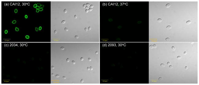
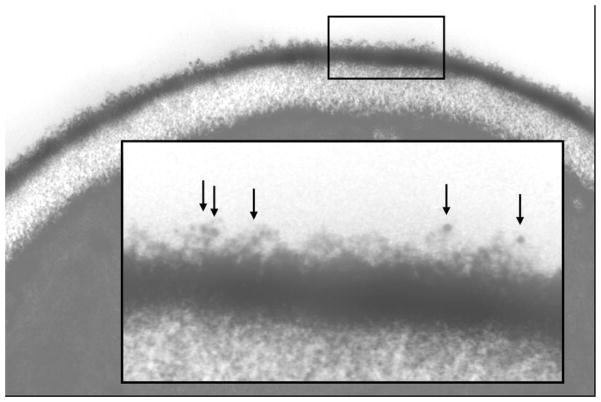
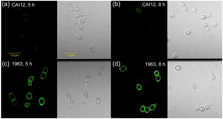
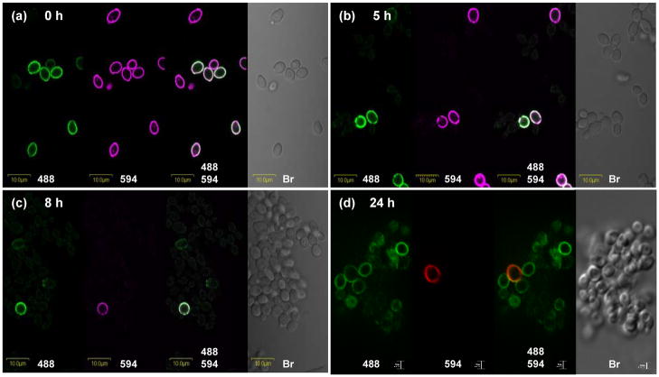
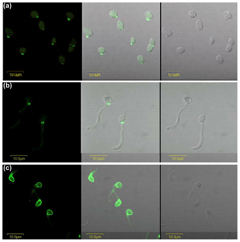
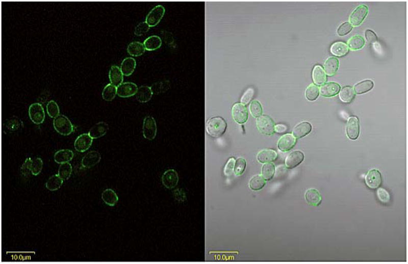
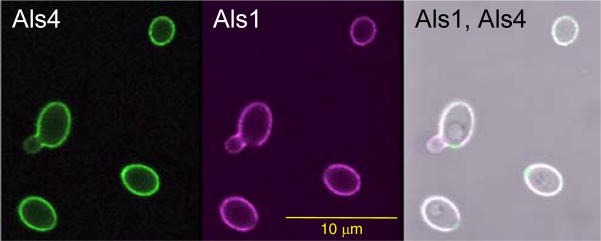
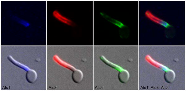
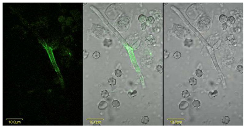
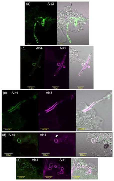
The Candida albicans agglutinin-like sequence (ALS) family encodes large cell surface glycoproteins that function in adhesion of the fungus to host and abiotic surfaces. Monoclonal antibodies (mAbs) specific for each Als protein were developed to study Als localization on the C. albicans surface. An anti-Als4 mAb demonstrated that Als4 covers the surface of yeast cells, with a greater abundance of Als4 on cells grown at 30 °C compared to 37 °C. On germ tubes, Als4 is localized in a restricted area proximal to the mother yeast. Immunolabeling with several anti-Als mAbs showed overlapping localization of Als1 and Als4 on yeast cells and Als1, Als3 and Als4 on germ tubes. Overlapping localization of Als proteins was also observed on yeast and hyphae recovered from mouse models of disseminated and oral candidiasis. Differences between Als localization in vivo and in vitro suggested changes in regulation of Als production in the host compared to the culture flask. Characterization with the anti-Als mAbs reveals the simultaneous presence and differences in relative abundance of Als proteins, creating an accurate image of Als representation and localization that can be used to guide conclusions regarding individual and collective Als protein function.


Figures

Fig. 1
Fig. 1
Immunolabeling of C. albicans yeasts with anti-Als4 MAb. Yeasts of C. albicans control strain CAI12 were grown in YPD for 16 h at either 30°C (a) or 37°C (b). C. albicans strains 2034 (als4Δ/als4Δ; c) and 2093 (als4Δ/als4Δ::ALS4; d) were grown at 30°C for 16 h. All cells were immunolabeled with anti-Als4 and a FITC-conjugated secondary antibody. Panels with a dark background have laser (488 nm) illumination; other panels are illuminated with white light. Als4 was detected on the surface of CAI12 cells grown at 30°C and just barely detected on yeasts grown at 37°C. Als4 was not detected on the surface of the deletion mutant strain 2034 (c) nor on the surface of the ALS4 reintegrant strain 2093 (d). Cells were imaged using an Olympus BX50 FluoView microscope.
Fig. 2
Fig. 2
Electron micrograph of a C. albicans yeast cell. Yeast cells of strain CAI12 were grown for 16 h at 30°C in YPD medium and immunolabeled with anti-Als4 MAb and a gold-conjugated secondary antibody, prior to fixation, embedding and sectioning. Gold particles are indicated by arrows shown in the higher-magnification inset. This methodology detects Als4 that was localized in the outermost flocculant layer of the C. albicans cell surface.
Fig. 3
Fig. 3
Anti-Als4 immunolabeling of yeast forms over the course of culture growth to determine if Als4 produced from the strain SC5314 ALS4 smaller allele can be detected on the C. albicans cell surface. C. albicans strains CAI12 (control; a and b) and 1963 (ALS4 overexpression; c and d) were grown in YPD at 30°C. At various time points, aliquots of culture were removed, fixed with paraformaldehyde and immunolabeled with anti-Als4. For CAI12, Als4 presence on the cell surface varies with stage of growth. Many inoculum cells were Als4-positive (see Fig. 1a), but became more rare in the culture as cell divisions produce Als4-negative yeasts. At 5 h post-inoculation (a), Als4-positive CAI12 cells became more difficult to detect. At 8 h post-inoculation, ALS4 expression began to increase in strain CAI12, producing Als4-positive daughter cells (b). In contrast, strain 1963 that produced Als4 constitutively under the control of the TPI1 promoter, showed a consistently strong presence on the surface of the yeast cells at 5 h (c) and at 8 h (d). Because the native ALS4 alleles in strain CAI12 were not transcribed actively at these time points, surface Als4 in strain 1963 was derived primarily from the PTPI1-ALS4 construct and supported the conclusion that Als4 produced from the smaller ALS4 allele was detectable on the C. albicans cell surface. Cells were imaged using an Olympus BX50 FluoView microscope.
Fig. 4
Fig. 4
Photomicrographs of Alexa 594-labeled and anti-Als4-labeled C. albicans yeast cells to assess the persistence of Als4 on the cell surface over the course of culture growth. Cells of strain CAI12 were grown in YPD medium at 30°C for 16 h and then covalently labeled with Alexa 594 (0 h; a). These cells were inoculated into fresh YPD medium and followed over time as they grew at 30°C and 200 rpm shaking. Cells were assessed by photomicroscopy with illumination at 488 nm (to detect the FITC-labeled secondary antibody that indicates Als4 presence), at 594 nm (to detect Alexa 594 that marks cells used in the culture inoculum) and white light (Br; bright field microscopy to visualize all cells present). Time points were taken at 5 h (b), 8 h (c) and 24 h (d). Autofluorescence of yeast cells results in faint outlines of cells at 488 nm (green); bleeding of the FITC signal into the 594 channel results in faint purple signals on cells at 594 nm. Signals from 594 nm are red in (d). Cells in panels a to c were imaged using an Olympus BX50 FluoView microscope. Cells in panel d were imaged using the Zeiss Axiovert 200M microscopy system (see Materials and methods).
Fig. 5
Fig. 5
Anti-Als4 immunolabeling of germ tubes. C. albicans CAI12 was grown in YPD medium for 16 h at 37°C, washed in DPBS, counted and transferred into pre-warmed RPMI 1640 medium at a cell density of 5 × 10⁶ cells ml−1. Cells were removed from the culture at 20 min (a) and 90 min (b), fixed in paraformaldehyde and immunolabeled with anti-Als4 and a FITC-conjugated secondary antibody. Cell-surface Als4 was visible as early as 20 min following inoculation of the RPMI 1640 culture. The restricted distribution of Als4 on germ tubes was visible at the later time point. Images were illuminated with laser only (488 nm; left panel), white light (right panel) or both (center panel). (c) Yeast cells were grown in YPD medium for 16 h at 30°C and transferred to RPMI 1640 medium at 37°C as described above. After 60 min, cells were fixed in paraformaldehyde and immunolabeled with anti-Als4. The mother yeast cells grown at 30°C had a strong Als4 presence on the surface that persisted as germ tube formation progressed. Localization of Als4 in a narrow band proximal to the mother yeast was similar to results for the 37°C-grown mother yeast. All cells were imaged using an Olympus BX50 FluoView microscope.
Fig. 6
Fig. 6
Anti-Als4 immunolabeling of C. tropicalis grown for 1 h in RPMI medium. C. tropicalis ATCC 201380 was grown for 16 h in YPD medium at 37°C and 200 rpm shaking. Cells were washed in DPBS, counted and inoculated into RPMI 1640 medium at a density of 5 × 10⁶ cells ml−1. After 1 h, cells were collected and fixed in paraformaldehyde. Fixed cells were immunolabeled with anti-Als4 and a FITC-conjugated secondary antibody. Cells were visualized with laser light (488 nm; left panel) or laser and white light (right panel). The Als4-positive signal indicated that the anti-Als4 MAb epitope was present on C. tropicalis. C. tropicalis cells grown for 16 h in YPD were also Als4-positive (data not shown). Cells were imaged using an Olympus FluoView microscope.
Fig. 7
Fig. 7
Overlapping localization of Als4 and Als1 on C. albicans yeast cells. CAI12 cells were grown for 16 h at 30°C, washed, counted and transferred to fresh YPD medium for 1 h at 30°C. Following fixation in paraformaldehyde, cells were immunolabeled with Alexa 488-labeled anti-Als4 and Alexa 594-labeled anti-Als1. Cells were imaged using an Olympus FluoView microscope. Als4 was visible on the surface of the yeast, with only a weak signal on the emerging bud. Als1 coated the surface of the yeast, with the exception of the bud scar. The weaker Als1 signal on the emerging bud matches previous observations for anti-Als1 immunolabeling of yeast (Coleman et al., 2010).
Fig. 8
Fig. 8
C. albicans germ tube labeled with anti-Als1, anti-Als3 and anti-Als4 MAbs to show overlapping localization of the three proteins. C. albicans germ tubes of strain CAI12 were grown in RPMI 1640 for 1 h. Cells were fixed in paraformaldehyde and immunolabeled with anti-Als4 and a FITC-conjugated secondary antibody, Alexa 633-labeled anti-Als1, and Alexa 593-labeled anti-Als3 as indicated under each image above. Cells were imaged using the Zeiss Axiovert 200M microscopy system (see Materials and methods). All images were illuminated with a mercury lamp. Standard filter sets for Cy5 (Alexa 633-Anti-Als1), rhodamine (Alexa 594-anti-Als3) and FITC (anti-Als4-FITC secondary antibody) were used. Upper images show individual or merged fluorescent signals while lower images include both fluorescent and bright field signals. The merged images showed that Als1, Als3 and Als4 were present simultaneously on the germ tube and had overlapping localizations.
Fig. 9
Fig. 9
A C. albicans hypha from murine kidney tissue immunolabeled with anti-Als4. A BALB/cByJ mouse was inoculated via the lateral tail vein with 5 × 10⁵ cells of C. albicans strain CAI12. At 28 h post-inoculation, the kidneys were removed, minced with a razor blade, and homogenized. A portion of the supernatant was treated with anti-Als4 and a FITC-conjugated secondary antibody. Cells were imaged using an Olympus BX50 FluoView microscope. Images above are illuminated with laser only (488 nm; left panel), white light (right panel) or both (center panel). Als4-positive cells were difficult to find, suggesting that they were rare in this model. Repetition of the experiment with other mice yielded the same result.
Fig. 10
Fig. 10
C. albicans hyphae and yeast recovered from a murine model of oral candidiasis and immunolabeled with anti-Als antibodies. Immunosuppressed mice were inoculated orally with C. albicans CAI12 (see Materials and methods). After 5 days of infection, mice were euthanized and the tongue and oral mucosa homogenized. C. albicans cells were collected using a cell strainer, washed, fixed in paraformaldehyde, and immunolabeled with anti-Als mAbs. Cells were imaged using an Olympus BX50 FluoView microscope. (a). Anti-Als3 labeled hyphae, but not yeast cells. The left panel was illuminated with laser (488 nm) and the right with laser and white light. (b and c). Anti-Als1 (center panels) labeled both hyphae and yeast. Als4-positive hyphae (left panels) were more difficult to find compared to anti-Als1-labeled hyphae. In contrast to the restricted localizations of Als4 and Als1 on hyphae from culture, the proteins were detected over greater lengths of the hyphae recovered from the mouse model. (d and e). Anti-Als4 (left) and anti-Als1 (second from left) immunolabeled yeast cells. In many instances, anti-Als4 and anti-Als1 labeled the entire surface of the same yeast cell. Previous in vitro analyses demonstrated that anti-Als1 labels the yeast cell surface, with the exception of the bud scar (Coleman et al., 2010). In vivo, cells with a lack of labeling at one pole were detected (see arrow in d) although far more cells appeared to have an entire surface covered with anti-Als1 (e).

References

    1. Argimon S, Wishart JA, Leng R, et al. Developmental regulation of an adhesin gene during cellular morphogenesis in the fungal pathogen Candida albicans. Eukaryot Cell. 2007;6:682–692. - PMC - PubMed
    1. Beucher B, Marot-Leblond A, Billaud-Nail S, Oh S-H, Hoyer LL, Robert R. Recognition of Candida albicans Als3 by the germ tube-specific monoclonal antibody 3D9.3. FEMS Immunol Med Microbiol. 2009;55:314–323. - PMC - PubMed
    1. Braun BR, van Het Hoog M, d’Enfert C, et al. A human-curated annotation of the Candida albicans genome. PLoS Genet. 2005;1:36–57. - PMC - PubMed
    1. Clancy CJ, Cheng S, Nguyen MH. Animal models of candidiasis. Methods Mol Biol. 2009;499:65–76. - PubMed
    1. Coleman DA, Oh S-H, Zhao X, Hoyer LL. Heterogeneous distribution of Candida albicans cell-surface antigens demonstrated with an Als1-specific monoclonal antibody. Microbiology. 2010;156:3645–3659. - PMC - PubMed

Publication types

MeSH terms