. 2012 Mar 1;21(5):1090-8.
doi: 10.1093/hmg/ddr538. Epub 2011 Nov 22.

Differential effects of a polyalanine tract expansion in Arx on neural development and gene expression

Affiliations

Differential effects of a polyalanine tract expansion in Arx on neural development and gene expression

MacLean Pancoast Nasrallah et al. Hum Mol Genet. .

Abstract

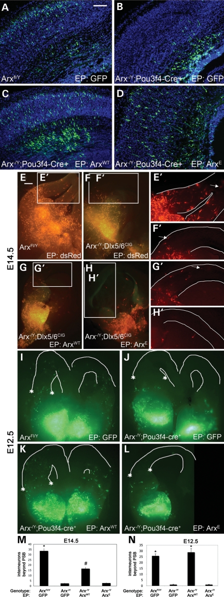
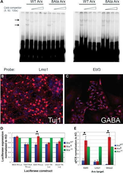
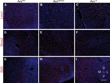
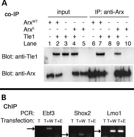
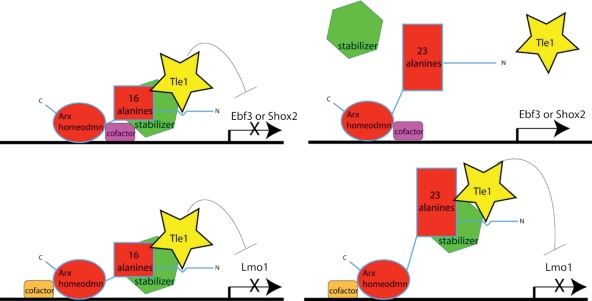
Polyalanine (poly-A) tracts exist in 494 annotated proteins; to date, expansions in these tracts have been associated with nine human diseases. The pathogenetic mechanism by which a poly-A tract results in these various human disorders remains uncertain. To understand the role of this mutation type, we investigated the change in functional properties of the transcription factor Arx when it has an expanded poly-A tract (Arx(E)), a mutation associated with infantile spasms and intellectual disabilities in humans. We found that although Arx(E) functions normally in the dorsal brain, its function in subpallial-derived populations of neurons is compromised. These contrasting functions are associated with the misregulation of Arx targets through the loss of the ability of Arx(E) to interact with the Arx cofactor Tle1. Our data demonstrate a novel mechanism for poly-A expansion diseases: the misregulation of a subset of target genes normally regulated by a transcription factor.


Figures

Figure 1.
Figure 1.
In utero electroporation (EP) of ArxWT, ArxE or control constructs into Arx-/Y;Pou3f4-cre+ (mutant) embryos compared with EP into wild-type littermates demonstrates that ArxE has a partial ability to rescue radial cell migration in the mutant mice; in contrast, slice electroporation of ArxE cannot rescue migration. (A) Wild-type embryos electroporated at E14.5 with a control vector expressing GFP were harvested at E18. Sections are counterstained with DAPI (blue). (B) An Arx-/Y;Pou3f4-cre+ brain electroporated with a control construct shows a defect in radial migration. (C and D) Arx-/Y; Pou3f4-cre+ embryos electroporated with ArxWT (C) or ArxE (D) show rescue of radial migration. Scale bar (upper right corner in A): 100 mm. (E) Normal NRCM is observed in E14.5 wild-type brains after EP of a control dsRed construct. White lines define the pial and ventricular surfaces. (F) In contrast, brains in which Arx is conditionally deleted in interneurons (Arx-/Y;Dlx5/6CIG) have loss of NRCM. Electroporation of ArxWT (G) but not ArxE (H) is able to partially rescue this migration defect. (E'–H') Detail of interneurons migrating in the pallium; the furthest-migrating interneuron is indicated with an arrow. Results were the same for Arx-/Y;Pou3f4-cre slice culture. (I) Control electroporation in wild-type E12.5 slice culture. (J) Loss of migration in E12.5 Arx-/Y;Pou3f4-cre slice culture. (K) NRCM rescue with ArxWT at E12.5. (L) Failure of rescue by ArxE at E12.5. A line drawn between the asterisks' define the location of the pallial–subpallial boundary (I–L, line not drawn so data are not obscured). (M) Counts in wild-type brains and both types of mutant brains electroporated with ArxWT were significantly different from all others (n > 10 for each genotype-rescue combination; *, #P < 0.05). A trend toward rescue was observed with electroporation of ArxWT but not ArxE into mutant brains. (N) Quantification of NRCM after E12.5 rescue shows ArxWT rescued NRCM at E12.5 (n > 7 for each genotype–rescue combination; *P < 0.05). No statistical differences were found between migration for Arx-/Y;Dlx5/6CIG and Arx-/Y;Pou3f4-Cre+ mutant mice; quantification includes both mutant lines. Each genotype/construct combination is represented by at least three brains from three different litters of each mutant line at each time point. PSB, pallial-subpallial boundary; error bars, SEM; scale bar (in A): 500 mm.
Figure 2.
Figure 2.
ArxE has not lost the ability to bind to Arx targets, but has lost the ability to repress a subset of them. (A) EMSA comparing ArxE and ArxWT binding to Lmo1 and Ebf3-binding sites shows that ArxE has not lost the ability to bind to Arx targets. Arrows indicate specific bands for Lmo1 and Ebf3, as determined by the loss of the bands upon addition of cold competitor of increasing concentrations. The lower band is most likely monomer, and the upper band is dimer, based on the absence of a band showing degraded protein (data not shown). Both upper and lower bands from four sets of experiments were quantitated using ImageJ and used to calculate Kd values. The Kd Arx/Kd ArxE was calculated for Ebf3 upper band (2.18) and lower band (1.65) as well as the upper (0.89) and lower band for Lmo1 (according to the protocol of (35). These data indicate both Arx and ArxE bind both targets with similar affinity. (BE) Measurement of Arx targets in differentiated ES cells shows loss of repression by ArxE for Ebf3 and Shox2 but not Lmo1. Three lines of ES cells were differentiated into neuronal populations: wild-type R1 (ArxWT), a line with Arx deleted (Arx-/Y), and a line engineered for Arx to have a poly-A expansion in the first alanine track (ArxE). (B) Tuj1 staining (DAPI counterstain) demonstrates extensive neuronal differentiation. (C) Many cells show GABAergic differentiation. (D) Luciferase assay shows that ArxE does not properly regulate Ebf3 expression. Luciferase data were normalized to Renilla expression. Data for the Arx-transfected data points are presented as percentage activation (±SEM) relative to empty vector-transfected cells (*P = 0.005). (E) Quantitative PCR data indicate the differential regulation of Arx targets in ES cells carrying the Arx poly-A expansion (±SEM, ArxWT versus ArxE for Ebf3 P = 0.004, for Shox2 P = 0.00009).
Figure 3.
Figure 3.
Aberrant expression of Ebf3 and Shox2, but not Lmo1, at E18 in Arx(GCG)7 mice. Wild-type and mutant E18 brain tissue were stained for Lmo1 (AC), Shox2 (DF) and Ebf3 (GI) in red, with blue DAPI counterstain. The three proteins show no expression (equal to repression) in wild-type tissue (A, D, G), but are expressed (not repressed) in Arx-/Y tissue (C, F, I). In Arx(GCG)7/Y tissue, Lmo1 continues to be repressed (B), but Shox2 and Ebf3 are not repressed, as seen in the Arx-/Y tissue (E and H). All images are from the ventral subpallium, with the ventricle to the left, and dorsal at the top, as indicated by the compass. Staining was performed for n = 4 for each genotype/antibody combination. Scale bar (see I): 120 mm.
Figure 4.
Figure 4.
Loss of cooperative binding of Tle1 and ArxE. (A) Tle1 co-immunoprecipitates less efficiently with ArxE than with ArxWT. Neuro-2a cells normally do not express Tle1 (input, lanes 1 and 5). After co-expression of Tle1 and ArxWT, Tle1 can be pulled down with Arx (IP, lane 7). The amount of Tle1 pulled down by ArxE is reduced (IP, lane 9). (B) After co-transfection with Tle1 and ArxWT, ChIP with anti-V5 antibody to pull down Tle1 shows that the three Arx target sequences upstream of Ebf3, Shox2 and Lmo1 are amplified (arrows indicate band size). However, after co-transfection of Tle1 and ArxE, only Lmo1 is amplified. T, Tle1; T + W, Tle1 and ArxWT; T + E, Tle1 and ArxE (also see Supplementary Material, Fig. S4C for quantitation).
Figure 5.
Figure 5.
Model of pathological mechanism resulting from expansion of first poly-A tract in Arx. The expansion results in a context-dependent loss of Tle recruitment to promoter sites. The context at the Ebf3 and Shox2 promoters does not allow Tle recruitment with ArxE (top right); however, the binding and activity of Tle are not perturbed in the case of Lmo1 (bottom right).

References

    1. Friocourt G., Parnavelas J.G. Mutations in ARX result in several defects involving GABAergic neurons. Front. Cell Neurosci. 2010;4:4. - PMC - PubMed
    1. Bienvenu T., Poirier K., Friocourt G., Bahi N., Beaumont D., Fauchereau F., Ben Jeema L., Zemni R., Vinet M.C., Francis F., et al. ARX, a novel Prd-class-homeobox gene highly expressed in the telencephalon, is mutated in X-linked mental retardation. Hum. Mol. Genet. 2002;11:981–991. - PubMed
    1. Guerrini R., Moro F., Kato M., Barkovich A.J., Shiihara T., McShane M.A., Hurst J., Loi M., Tohyama J., Norci V., et al. Expansion of the first PolyA tract of ARX causes infantile spasms and status dystonicus. Neurology. 2007;69:427–433. - PubMed
    1. Bauer P.O., Nukina N. The pathogenic mechanisms of polyglutamine diseases and current therapeutic strategies. J. Neurochem. 2009;110:1737–1765. - PubMed
    1. Messaed C., Rouleau G.A. Molecular mechanisms underlying polyalanine diseases. Neurobiol. Dis. 2009;34:397–405. - PubMed

Publication types

MeSH terms