. 2011 Nov 22;2(6):e00257-11.
doi: 10.1128/mBio.00257-11. Print 2011.

Cellular architecture mediates DivIVA ultrastructure and regulates min activity in Bacillus subtilis

Affiliations

Cellular architecture mediates DivIVA ultrastructure and regulates min activity in Bacillus subtilis

Prahathees Eswaramoorthy et al. mBio. .

Abstract

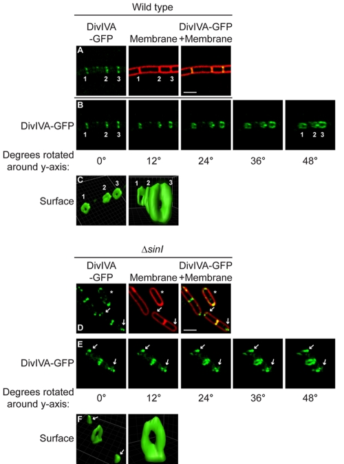
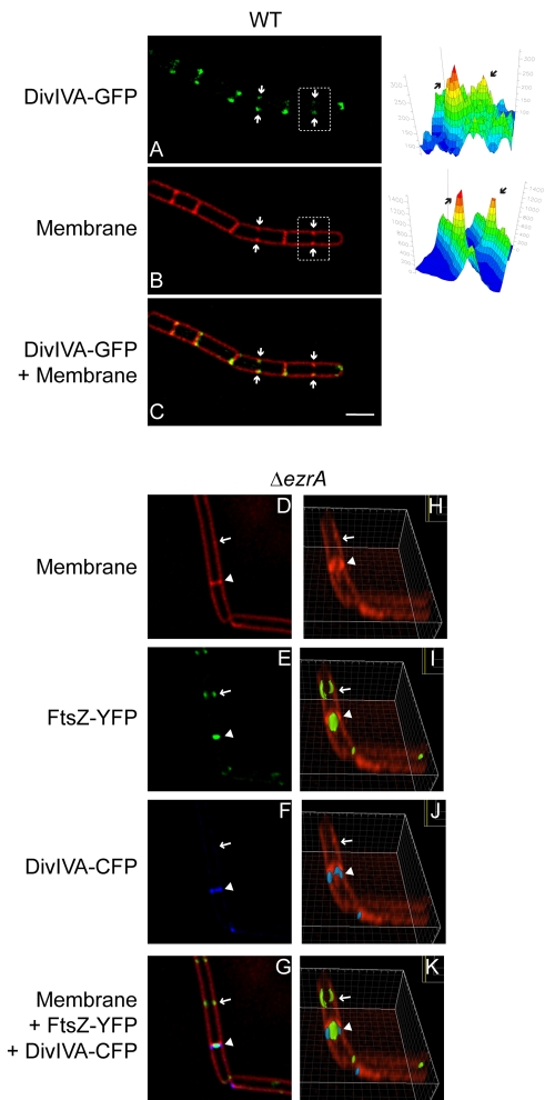
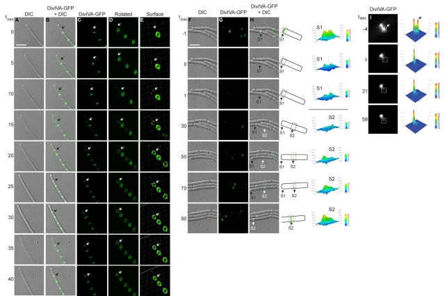
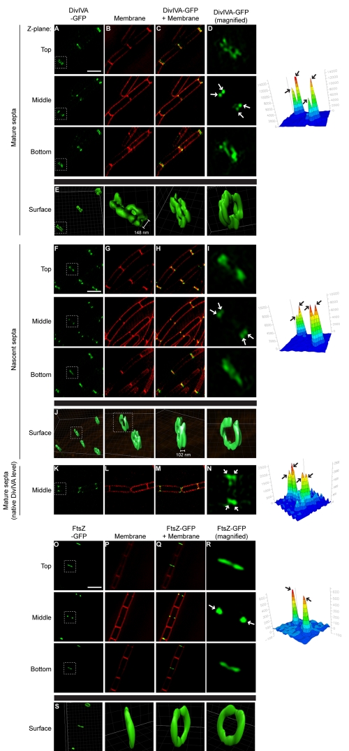
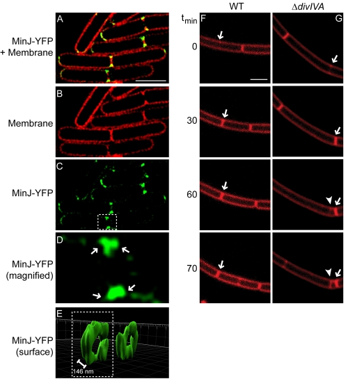
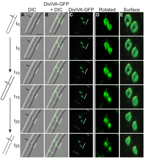
The assembly of the cell division machinery at midcell is a critical step of cytokinesis. Many rod-shaped bacteria position septa using nucleoid occlusion, which prevents division over the chromosome, and the Min system, which prevents division near the poles. Here we examined the in vivo assembly of the Bacillus subtilis MinCD targeting proteins DivIVA, a peripheral membrane protein that preferentially localizes to negatively curved membranes and resembles eukaryotic tropomyosins, and MinJ, which recruits MinCD to DivIVA. We used structured illumination microscopy to demonstrate that both DivIVA and MinJ localize as double rings that flank the septum and first appear early in septal biosynthesis. The subsequent recruitment of MinCD to these double rings would separate the Min proteins from their target, FtsZ, spatially regulating Min activity and allowing continued cell division. Curvature-based localization would also provide temporal regulation, since DivIVA and the Min proteins would localize to midcell after the onset of division. We use time-lapse microscopy and fluorescence recovery after photobleaching to demonstrate that DivIVA rings are highly stable and are constructed from newly synthesized DivIVA molecules. After cell division, DivIVA rings appear to collapse into patches at the rounded cell poles of separated cells, with little or no incorporation of newly synthesized subunits. Thus, changes in cell architecture mediate both the initial recruitment of DivIVA to sites of cell division and the subsequent collapse of these rings into patches (or rings of smaller diameter), while curvature-based localization of DivIVA spatially and temporally regulates Min activity.

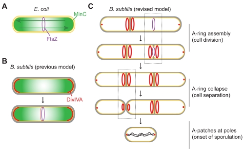
Importance: The Min systems of Escherichia coli and Bacillus subtilis both inhibit FtsZ assembly, but one key difference between these two species is that whereas the E. coli Min proteins localize to the poles, the B. subtilis proteins localize to nascent division sites by interaction with DivIVA and MinJ. It is unclear how MinC activity at midcell is regulated to prevent it from interfering with FtsZ engaged in medial cell division. We used superresolution microscopy to demonstrate that DivIVA and MinJ, which localize MinCD, assemble double rings that flank active division sites and septa. This curvature-based localization mechanism holds MinCD away from the FtsZ ring at midcell, and we propose that this spatial organization is the primary mechanism by which MinC activity is regulated to allow division at midcell. Curvature-based localization also conveys temporal regulation, since it ensures that MinC localizes after the onset of division.


Figures

FIG 1
FIG 1
Ultrastructure of DivIVA rings at division septa and DivIVA patches at hemispherical poles. (A) Localization of DivIVA-GFP (green) in wild-type B. subtilis growing as a chain of cells that have completed cytokinesis but not cell separation (strain KR515, left). The center panel shows a membrane visualized with the fluorescent dye FM4-64 (red). The right panel shows overlay of fluorescence from DivIVA-GFP and FM4-64; regions of fluorescence overlap are yellow. (B and E) Rotation of the images in panels A and D, respectively, around the y axis (degrees rotated are indicated below each panel). Two older septa are labeled “1” and “2,” and a nascent division septum is labeled “3.” (C and F) DivIVA-GFP fluorescence in panels B and E, respectively, represented as a three-dimensional surface. (D) Localization of DivIVA-GFP in ΔsinI cells that grow as individual cells during exponential phase (strain KR528). At center and right, membrane stain and overlay are shown. Arrows indicate DivIVA patches at the poles. A cell that had not elaborated a septum is marked with an asterisk. Scale bar, 2 µm. Strains are described in Materials and Methods.
FIG 2
FIG 2
DivIVA localizes to septa at the onset of membrane constriction. (A) DivIVA-GFP localization in strain KR515. (B) Membrane visualized with FM4-64. (C) Overlay of DivIVA-GFP and FM4-64. Arrows indicate invaginating membranes at nascent cell division sites. Quantification of fluorescence intensity from FM4-64 and DivIVA-GFP for one incomplete septum (white box) is on the right (arrows). (D to G) Localization of DivIVA-CFP and FtsZ-YFP in the ΔezrA strain (PE99). (D) Membrane visualized with FM4-64. (E) FtsZ-YFP. (F) DivIVA-CFP. (G) Overlay of FM4-64, FtsZ-YFP, and DivIVA-CFP. (H to K) Rotation of the images in (D to G), respectively, around the x axis. The arrow indicates location of an assembled but nonfunctional FtsZ ring; the arrowhead indicates an actively constricting FtsZ ring. Scale bar, 2 µm.
FIG 3
FIG 3
Assembly of stable A rings at septa. (A to E) Time-lapse micrographs of DivIVA-GFP localization in strain KR541. (A) Differential interference contrast (DIC). (B) Overlay, DivIVA-GFP and DIC. (C) DivIVA-GFP. (D) Images in panel C rotated to view A rings. (E) DivIVA-GFP fluorescence intensity in panel D, represented as a three-dimensional surface. Time (minutes) is shown at left. The arrow indicates the site of a nascent A ring assembling. (F to H) DIC, DivIVA-GFP, and overlay in which fluorescence recovery was monitored after photobleaching a field of cells (strain KR528). Time (minutes) before and after photobleaching is shown at left. “S1” is a preexisting septum, and “S2” is a septum formed after photobleaching. A schematic representation of DivIVA-GFP localization in panel H at either S1 or S2 is shown to the right, along with quantification of fluorescence intensities of S1 and S2 before and after photobleaching. Scale bar, 5 µm. (I) Fluorescence recovery was monitored after photobleaching half of an A ring (white box). Time (seconds) before or after photobleaching is indicated at left, and quantification of fluorescence intensity is at right.
FIG 4
FIG 4
Adjacent A rings at division septa. DivIVA-GFP localization (strain KR541, produced under control of an IPTG-inducible promoter) at mature (A to E) or nascent (F to J) division septa, DivIVA-GFP localization (strain KR515, produced at native levels) at mature division septa (K to N), and FtsZ-GFP localization (strain AD3007) (O to S) viewed by 3D-SIM at three Z planes (indicated as “Top,” “Middle,” or “Bottom”). (B, G, L, and P) Membranes in panels A, F, K, and O, respectively, visualized using FM4-64. (C, H, M, and Q) Overlay of membrane stain and GFP fluorescence. (D, I, N, and R) Magnification of one septum (white box) in panels A, F, K, and O, respectively. (E, J, and S) GFP fluorescence for selected septa in panels A, F, and O, represented as a three-dimensional surface. Separation between A rings indicated is the average distance between regions of peak fluorescence (n = 24 for doublet A rings at mature septa, ±20 nm; n = 8 for doublet A rings at incomplete septa, ±14 nm). GFP fluorescence intensity for the selected septa in panels D, I, N, and R at the intermediate Z plane was quantified and is shown on the right to highlight the number of foci (arrows). Scale bars, 2 µm.
FIG 5
FIG 5
A rings mediate fidelity of cell division by recruiting MinJ to both sides of active division sites. (A) Overlay of FM4-64 and MinJ-YFP fluorescence, viewed by 3D-SIM at an intermediate Z plane (strain DS3609). (B) Membrane stain. (C) MinJ-YFP. (D) Magnification of one septum (white box) in panel C. (E) YFP fluorescence for selected septa in panel C, represented as a three-dimensional surface. Separation between MinJ rings indicated is the average distance between regions of peak fluorescence, ±16 nm (n = 8). Time-lapse images of dividing wild-type (WT) (F) (strain PY79) or ΔdivIVA (G) (strain KR546) cells were visualized using FM4-64. Time (minutes) after the initiation of the experiment is indicated on the left. Arrows indicate recently completed septa; arrowhead indicates a mispositioned septum. Scale bars, 2 µm.
FIG 6
FIG 6
Assembly of A patches at hemispherical poles. (A to E) Time-lapse micrographs of DivIVA-GFP localization in strain KR541. (A) DIC image. Arrow indicates a septum whose architecture is represented schematically on the left. (B) Overlay of DivIVA-GFP and DIC. (C) DivIVA-GFP. (D) Indicated region in panel C, magnified and rotated. (E) DivIVA-GFP fluorescence intensity from the deconvolved images in panel D, represented as a three-dimensional surface. Scale bar, 2 µm.
FIG 7
FIG 7
Model for the architecture-driven assembly of A rings and A patches. (A) Role of the Min system in establishing the midcell in Escherichia coli. An E. coli cell initiating cell division is depicted (inner and outer membranes, yellow; FtsZ ring, purple; MinC, green), wherein the pole-to-pole oscillation of MinC (green arrows) results in a time-averaged MinC concentration gradient that is minimal at midcell, where FtsZ polymerizes. (B) Previous model of the Min system in B. subtilis. A single B. subtilis cell is depicted (plasma membrane, yellow; cell wall, gray). Analogous to the E. coli model, DivIVA (red) was previously thought to primarily localize to the hemispherical poles, where it sequestered MinC in order to create a static MinC concentration gradient that is minimal at midcell. The fainter localization of DivIVA and MinC to midcell was proposed to occur at a time at which the FtsZ ring was resistant to MinC activity, thereby allowing continued division. (C) Revised model of the role of the Min system in B. subtilis. A chain of B. subtilis cells is depicted in which DivIVA localizes at the onset of membrane constriction and assembles into doublet A rings that recruit MinC (not shown for simplicity) to two rings that flank the active division site. This spatially separates MinC and FtsZ, allowing continued cell division, and mediates the transient localization of MinC adjacent to the newly formed septum, where it can prevent formation of an aberrantly positioned division septum. We therefore propose that the curvature-based DivIVA localization mechanism conveys both temporal and spatial regulation on MinC, preventing it from acting on FtsZ at the active division site. As chains of cells separate, A rings collapse into patches, or perhaps ring with small central openings, as flat septa transform into hemispherical poles.

Similar articles

Cited by

References

    1. Berg CA. 2005. The Drosophila shell game: patterning genes and morphological change. Trends Genet. 21:346–355 - PubMed
    1. Margolis HC, Beniash E, Fowler CE. 2006. Role of macromolecular assembly of enamel matrix proteins in enamel formation. J. Dent. Res. 85:775–793 - PubMed
    1. Leiman PG, Kanamaru S, Mesyanzhinov VV, Arisaka F, Rossmann MG. 2003. Structure and morphogenesis of bacteriophage T4. Cell. Mol. Life Sci. 60:2356–2370 - PMC - PubMed
    1. Shapiro L, McAdams HH, Losick R. 2009. Why and how bacteria localize proteins. Science 326:1225–1228 - PMC - PubMed
    1. Rudner DZ, Losick R. 2010. Protein subcellular localization in bacteria. Cold Spring Harb. Perspect. Biol. 2:a000307 - PMC - PubMed

Publication types

MeSH terms