. 2012 Mar;60(3):229-42.
doi: 10.1369/0022155411431734. Epub 2011 Nov 21.

Age-related accumulation of non-heme ferric and ferrous iron in mouse ovarian stroma visualized by sensitive non-heme iron histochemistry

Affiliations

Age-related accumulation of non-heme ferric and ferrous iron in mouse ovarian stroma visualized by sensitive non-heme iron histochemistry

Yoshiya Asano. J Histochem Cytochem. 2012 Mar.

Abstract

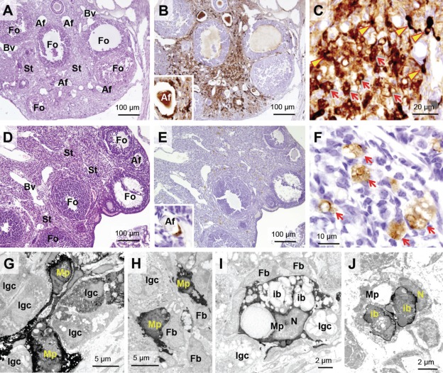
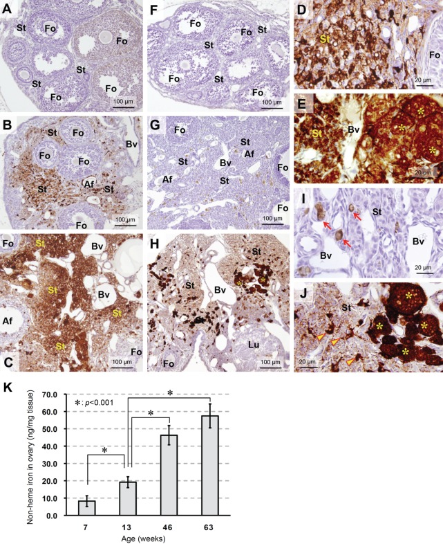
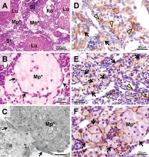
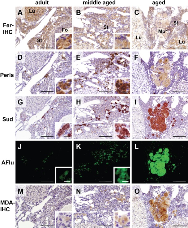
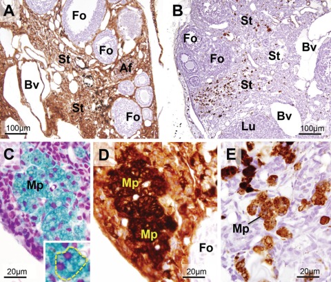
Sensitive non-heme iron histochemistry--namely, the perfusion-Perls method and perfusion-Turnbull method--was applied to study the distribution and age-related accumulation of non-heme ferric iron and ferrous iron in mouse ovary. Light and electron microscopic studies revealed that non-heme ferric iron is distributed predominantly in stromal tissue, especially in macrophages. By contrast, the distribution of non-heme ferrous iron was restricted to a few ovoid macrophages. Aged ovaries exhibited remarkable non-heme iron accumulation in all stromal cells. In particular, non-heme ferrous iron level was increased in stromal tissue, suggestive of increased levels of redox-active iron, which can promote oxidative stress. Moreover, intense localization of both non-heme ferric and ferrous iron was observed in aggregated large stromal cells that were then characterized as ceroid-laden enlarged macrophages with frothy cytoplasm. Intraperitoneal iron overload in adult mice resulted in non-heme iron deposition in the entire stroma and generation of enlarged macrophages, suggesting that excessive iron accumulation induced macrophage morphological changes. The data indicated that non-heme iron accumulation in ovarian stromal tissue may be related to aging of the ovary due to increasing oxidative stress.


Conflict of interest statement

The authors declared no potential conflicts of interest with respect to the authorship and/or publication of this article.

Figures

Figure 1.
Figure 1.
Distribution of NHF[III] and NHF[II] in ovarian tissue of adult mice visualized by sensitive non-heme iron histochemistries. (A–C) Visualization of NHF[III] in adult mice (14 weeks old) by perfusion-Perls method (PPIrH) under light microscope. (A) PPIrH and hematoxylin and eosin (HE) staining. (B, C) PPIrH and 3,3′-diaminobenzidine·4HCl (DAB) intensification. (A, B) NHF[III]-positive staining (brown color) was mainly observed in stroma (St). Around atretic follicles (Af), intense staining was observed by localization of macrophages with NHF[III] positive (B, inset). Granulosa cells of growing follicles (Fo) were negative or palely positive. Follicular fluid was weakly stained. (C) Macrophages with thin-branched shape (thin-branched macrophages) were densely stained (arrowheads). The other stained cells were macrophages with an ovoid shape or interstitial gland cells (arrows). (D–F) Visualization of NHF[II] in adult mice (14 weeks old) by perfusion Turnbull method (PTIrH). (D) PTIrH and HE staining. (E, F) PTIrH and DAB intensification. NHF[II]-positive staining (brown color) was observed in only a few cells in the stroma and atretic follicles (inset). No staining was observed in follicles. (F) Macrophages with ovoid shape and vacuoles (ovoid macrophages) were stained (arrows). (G–I) Electron microscopy of ovarian stroma stained using PPIrH. The staining of NHF[III] (high electron density) was strongly observed in thin-branched macrophages (G and H, Mp). Some of interstitial gland cells were slightly stained (G and H, Igc). Fibroblasts were negative (H and I, Fb). (I) Ovoid macrophages (Mp) were densely stained by PPIrH. The staining was observed in the cytosol, rather than inclusion bodies (ib) or lipid droplets. (J) Electron microscopy of stromal ovoid macrophages (Mp) stained by PTIrH. Intense staining of NHF[II] with high electron density was observed at peripheral parts of inclusion bodies (ib). N, nucleus; Bv, blood vessels. For sensitive non-heme iron histochemistries in light microscopy, at least four female mice in each age group were used, with the representative data shown.
Figure 2.
Figure 2.
Change of NHF[III] and NHF[II] distribution in ovarian tissue as aging. (A–C) Change of NHF[III] distribution as aging visualized by perfusion-Perls method (PPIrH) and 3,3′-diaminobenzidine·4HCl (DAB) intensification. (F–H) Change of NHF[II] distribution as aging visualized by perfusion Turnbull method (PTIrH) and DAB intensification. (A, F) Ovary from immature mouse (4 weeks old). (A) No staining of NHF[III] was found in the stroma (St). In some follicles (Fo), weak staining was observed in granulosa cells. (F) No staining of NHF[II] was found. (B, G) Ovary from adult mouse (14 weeks old). In the expanded stroma, distribution of NHF[III] and NHF[II] was observed as in Figure 1B,E. (C, H) Ovary from aged mouse (52 weeks old). (C) Stroma (St) further expanded with aging. The density of NHF[III] staining was higher than that of adult ovary, suggesting accumulation of non-heme iron. Growing follicles did not show increase of staining compared to adult ovary. Af, atretic follicle; Bv, blood vessels. (H) Increase of NHF[II] staining was observed in entire of stroma (St), including enlarged macrophages with extremely dense staining (asterisks). Growing follicles (Fo) and corpora lutea (Lu) were not stained. (D, E) Ovarian stroma of adult mouse (D) and aged mouse (E) stained by PPIrH. In aged mouse (E), the staining was intense and homologous among stromal cells except for blood vessels. Clusters of large round cells, characterized as macrophages by F4/80 immunostaining (Fig. 3), were intensely stained (asterisks). (I, J) Ovarian stroma of adult mouse (I) and aged mouse (J) stained by PTIrH. In the aged ovary (J), NHF[II] staining was increased over the stroma, compared to adult ovary (I). Clusters of large and round macrophages were intensely stained (asterisks). Also, positively stained thin-branched macrophages increased (arrowheads). (K) Quantitative analysis of non-heme iron in ovarian tissue from immature mice (7 weeks old), adult mice (13 weeks old), and aged mice (46 and 63 weeks old). Results are mean ± SD (n=4). The amount of non-heme iron in ovarian tissue clearly increased with age (p<0.001 in 13 weeks old vs 7, 46, or 63 weeks old).
Figure 3.
Figure 3.
Enlarged macrophages in ovarian stroma of aged mice. (A, B) Hematoxylin and eosin (HE) staining. Enlarged macrophages (Mp*) located in stroma as clusters. Their cytoplasm filled with vacuoles was palely stained with eosin. Multiple nuclei and incomplete cellular borders were observed, suggesting fusion of the cells. The clusters were surrounded by thin and flat cells (arrows). (C) Electron microscopy of enlarged macrophages (Mp*) with customary electron staining. Cytoplasm was filled with polymorphic inclusion bodies and lipid droplets. Granules with high electron density, probably lysosomes, were also observed. The flat cells surrounding enlarged macrophages were considered mechanically pressured fibroblasts according to their morphological characteristics. (D–F) Ovarian stroma stained with F4/80 immunostaining as a marker for mouse macrophages. (D) Ovary of an adult mouse (14 weeks old). The staining was observed at the surface of thin-branched macrophages (arrowheads) and ovoid macrophages (arrows). (E) Ovary of a middle-aged mouse (34 weeks old). The number of ovoid macrophages with F4/80 immunostaining was increased in stroma (arrows). (F) Ovary of an aged mouse (52 weeks old). The immunostaining was observed at the periphery of enlarged macrophages (arrows). These observations suggest that the enlarged macrophages are derived from ovoid macrophages in the stroma of adult and middle-aged ovaries. St, stroma; Fo, follicles; Lu, corpus luteum.
Figure 4.
Figure 4.
Co-localization of iron and ceroid in ovoid macrophages and enlarged macrophages. Serial sections of ovaries from adult mouse (14 weeks old, left column), middle-aged mouse (34 weeks old, center column), and aged mouse (52 weeks old, right column) were stained by ferritin immunohistochemistry (Fer-IHC, A–C), customary Perls method (Perls, D–F), Sudan III staining (Sud, G–I), and malondialdehyde immunohistochemistry (MDA-IHC, M–O). Autofluorescence was also observed (AFlu, J–L). Ferritin immunohistochemistry demonstrated the staining in almost every part of ovarian tissue of adult (A) and middle-aged (B) ovary. Ovoid macrophages were also positively stained (insets of A and B), although only pale staining was observed in enlarged macrophages in aged ovary (C, Mp*). Customary Perls method to adjoining sections showed intense staining of NHF[III] in ovoid macrophages in adult and middle-aged ovary (D and E, insets). In aged ovary, clusters of enlarged macrophages were weakly stained (F). Note that these cells were densely stained by perfusion-Perls method (PPIrH) and perfusion Turnbull method (PTIrH) (Fig. 2E,J). On the other hand, ovoid macrophages in adult (G and J) and middle-aged (H and K) ovary and enlarged macrophages in aged ovary (I and L) were intensely stained by Sudan III and emitted strong autofluorescence by 480 nm excitation light, demonstrating presence of ceroid. MDA immunohistochemistry also showed the staining of ovoid macrophages in adult ovary (M, inset), middle-aged ovary (N, inset), and enlarged macrophages in aged ovary (O), with gradual increase of staining intensity with age, suggesting accumulation of oxidative stress. Fo, follicle; Lu, corpus luteum; St, stroma. Scale bar: 100 µm (inset: 10 µm).
Figure 5.
Figure 5.
Distribution of NHF[III] and NHF[II] in ovarian tissue of iron-overloaded adult mouse. (A) Distribution of NHF[III] visualized by perfusion-Perls method (PPIrH) and 3,3′-diaminobenzidine·4HCl (DAB) intensification. Intense staining was observed in the entire stroma and theca externa except for growing follicles. (B) Distribution of NHF[II] visualized by perfusion Turnbull method (PTIrH) and DAB intensification. The increased staining was restricted in the stromal ovoid macrophages (brown spots). (C, D) The cluster of enlarged macrophages (Mp) in stroma of iron-overloaded ovary. (C) PPIrH and hematoxylin and eosin (HE) staining. (D) PPIrH and DAB intensification. These enlarged macrophages showed dense staining of NHF[III] (blue color in C and brown color in D), frothy cytoplasm, and multiple nuclei (inset of C; nuclei were stained by hematoxylin). (E) Stroma in iron-overloaded ovary stained by PTIrH with DAB intensification. Enlarged macrophages (Mp) were NHF[II] positive. Fo, follicle; Af, atretic follicles; Lu, corpus luteum; St, stroma; Bv, blood vessels.

References

    1. Agarwal A, Gupta S, Sharma RK. 2005. Role of oxidative stress in female reproduction. Reprod Biol Endocrinol. 3:28. - PMC - PubMed
    1. Agarwal A, Gupta S, Sikka S. 2006. The role of free radicals and antioxidants in reproduction. Curr Opin Obstet Gynecol. 18:325–332 - PubMed
    1. Aleshire SL, Osteen KG, Maxson WS, Entman SS, Bradley CA, Parl FF. 1989. Localization of transferrin and its receptor in ovarian follicular cells: morphologic studies in relation to follicular development. Fertil Steril. 51:444–449 - PubMed
    1. Asano Y, Meguro R, Odagiri S, Li C, Iwatsuki H, Shoumura K. 2006. Visualization of non-heme ferric and ferrous iron by highly sensitive non-heme iron histochemistry in the stress-induced acute gastric lesions in the rat. Histochem Cell Biol. 125:515–525 - PubMed
    1. Behrman HR, Kodaman PH, Preston SL, Gao S. 2001. Oxidative stress and the ovary. J Soc Gynecol Investig. 8(1 Suppl Proceedings):40–42 - PubMed

Publication types

MeSH terms