. 2011 Oct 23;3(12):925-31.
doi: 10.1038/nchem.1174.

Cytocompatible click-based hydrogels with dynamically tunable properties through orthogonal photoconjugation and photocleavage reactions

Affiliations

Cytocompatible click-based hydrogels with dynamically tunable properties through orthogonal photoconjugation and photocleavage reactions

Cole A DeForest et al. Nat Chem. .

Abstract

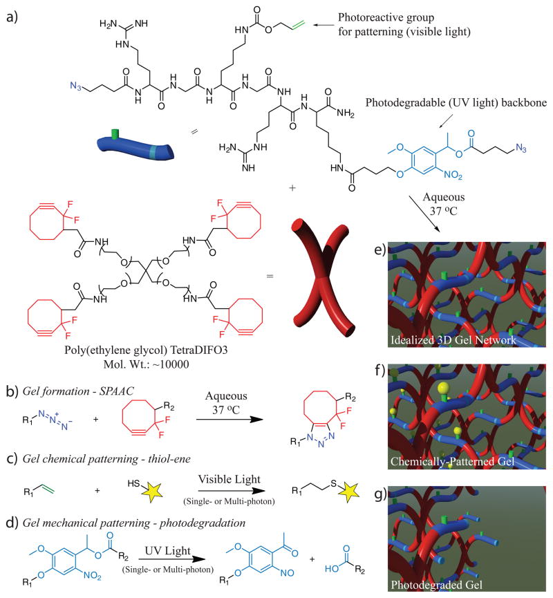
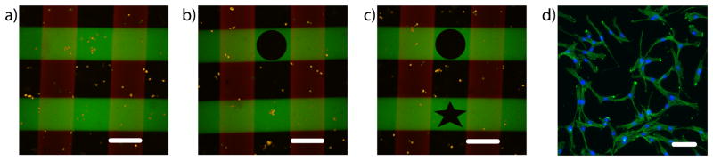
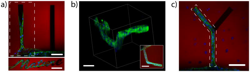
To provide insight into how cells receive information from their external surroundings, synthetic hydrogels have emerged as systems for assaying cell function in well-defined microenvironments where single cues can be introduced and subsequent effects individually elucidated. However, as answers to more complex biological questions continue to be sought, advanced material systems are needed that allow dynamic alteration of the three-dimensional cellular environment with orthogonal reactions that enable multiple levels of control of biochemical and biomechanical signals. Here, we seek to synthesize one such three-dimensional culture system using cytocompatible and wavelength-specific photochemical reactions to create hydrogels that allow orthogonal and dynamic control of material properties through independent spatiotemporally regulated photocleavage of crosslinks and photoconjugation of pendant functionalities. The results demonstrate the versatile nature of the chemistry to create programmable niches to study and direct cell function by modifying the local hydrogel environment.


Conflict of interest statement

Competing financial interests

The authors declare that they have no competing financial interests.

Figures

Figure 1
Figure 1. Synthesis, photocoupling, and photodegradation for tuning chemical and physical properties of click-based hydrogels
(a) Click-functionalized macromolecular precursors (i.e., PEG-tetraDIFO3 and bis(azide)-functionalized polypeptides) form a 3D ideal hydrogel structure via a step-growth polymerization mechanism by the (b) SPAAC reaction. (c) In the presence of visible light (λ = 490 – 650 nm or 860 nm), thiol-containing biomolecules are covalently affixed to pendant vinyl functionalities throughout the hydrogel network via the thiol-ene reaction. (d) A nitrobenzyl ether moiety within the backbone of the polymer network undergoes photocleavage in the presence of single or multi-photon UV light (λ = 365 nm or 740 nm) that results in photodegradation of the network. Schematics of the formed SPAAC-based idealized gel, network post thiol-ene functionalization, and material post photodegradation are found in (eg), respectively.
Figure 2
Figure 2. Biochemical patterning within preformed click hydrogels using visible light
Upon swelling thiol-containing biomolecules into preformed gels, pendant functionalities are affixed to the hydrogel backbone via the thiol-ene reaction upon exposure to visible light (λ = 490 – 650 nm). (a) The final patterned concentration of a fluorescent RGD peptide (AF488-AhxRGDSC-NH2) depends on the amount of photoinitiator present (2.5, 5, and 10 mM Eosin Y), as well as the exposure time to visible light (0 – 2 min, 10 mW cm−2). (b) The network functionalization with pendant fluorescently-labeled peptides is confined to user-defined regions within preformed gels using photolithography (0.5, 1, or 2 min exposure with increased patterning concentration for increased exposure time, 10 mW cm−2, 10 μM eosin Y). (c) The photocoupling reaction is controlled in 3D by rastering the focal point of multi-photon laser light (λ = 860 nm) over defined volumes within the gel, affording micron-scale resolution in all spatial dimensions. Additionally, the patterning process can be repeated many times to introduce multiple biochemical cues within the same network, as demonstrated by the red- and green- labeled patterned peptides within the same gel. Images represent confocal projections and 3D renderings. Scale bar = 400 μm for (b) and 100 μm for (c).
Figure 3
Figure 3. Biophysical patterning within preformed click hydrogels using UV light
In the presence of UV light (λ = 365 nm), photolabile functionalities within the hydrogel crosslinks undergo an irreversible cleavage, thereby decreasing the total network connectivity, resulting in local material degradation and removal of the fluorescent hydrogel material. (a) In optically-thick samples, the depth of photodegradation is directly related to the incident light intensity (5, 10 and 20 mW cm−2), as well as the exposure time to visible light (0 – 45 min). (b) Using mask-based photolithographic techniques, network degradation was confined to user-defined regions within fluorescently-labeled gels (10, 15, 20, 30, and 45 min, feature height increasing with exposure time), as measured by profilometry. (c) The photodegradation reaction is controlled in 3D with micron-scale resolution in all dimensions using focused multi-photon laser light (λ = 740 nm). Images represent confocal projections and 3D renderings. Scale bar = 400 μm for (b) and 100 μm for (c).
Figure 4
Figure 4. Orthogonality of photocoupling and photodegration reactions
(a) The peak absorbance for the photoinitiator (red) and the photolabile group (blue) is well separated (~520 and ~350 nm, respectively), thus enabling photocoupling and photodegradation reactions to be performed independently from one another using different light sources (illustrated with colored bars). (b) NMR studies indicate that the photodegradable moiety cleaves readily in the presence of UV light (λ = 365 nm, 10 mW cm−2), but remains intact when exposed to the visible light used to initiate the photocoupling reaction (λ = 490 – 650 nm, 10 mW cm−2). (c) Multi-photon visible light was first used to couple a fluorescently-labeled peptide within the center of the hydrogel in a user-defined 3D pattern (top, buffalo), and the network was subsequently degraded locally with multi-photon UV light, thereby removing the peptide from selected regions (bottom, CU and horn). (d) Brightfield microscopy confirms that photocleavage is confined only to user-defined locations within the gel, and that the photocoupling light conditions do not give rise to undesired degradation. Images in (c) represent 3D renderings of confocal z-stacks. Scale bars = 100 μm.
Figure 5
Figure 5. Culture and recovery of hMSCs from hydrogel microenvironments
CellTracker Orange-labeled human mesenchymal stem cells (hMSCs) were encapsulated within the click hydrogel formulation at 5 × 10⁶ cells mL−1. (a) 24 hours post encapsulation, perpendicular 200 μm wide lines of ~1 mM RGD and PHSRN were patterned throughout the hydrogel via thiol-ene photocoupling to create an array of four distinct biochemical conditions (no cue, RGD, PHSRN, RGD and PHSRN). (b) 4 hours later, channels of user-defined shape (cylindrical) were eroded down from the surface of the hydrogel to capture entrapped cells exposed to a specific cue. (c) This process was repeated 1 hour later to release entrapped cells within a different location of the material and a different shape (star-shaped cylinder). (d) The released cells were isolated by centrifugation, cultured in a 96-well plate for 48 hours, and their cytoskeleton was visualized with a fluorescent phalloidin. (a–c) RGD is shown in green, PHSRN red, and hMSCs orange. Images represent single confocal slices within the 3D gel. (d) F-actin is shown in green, nuclei blue. Image represents inverted fluorescence micrograph. Scale bars = 200 μm in (a–c), 50 μm in (d).
Figure 6
Figure 6. Directed 3D cell motility within patterned hydrogels
(a) A fibrin clot containing 3T3 fibroblasts was encapsulated within the click hydrogel formulation. Chemical channels of RGD, a cell-adhesive fibronectin motif, as well as physical channels of user-defined shape were created radially out of the roughly spherical clot. The combination of having physical space to spread as well as chemical moieties to bind to were found to be required for collective cell migration. By day 10, cells were found to migrate only down the physical channel that was functionalized with RGD. (b) By creating 3D functionalized channels, cell outgrowth was controlled in all three spatial dimensions, with the image inset illustrating a top-down projection. (c) The outgrowth of 3T3 fibroblast cells was controlled in the presence of encapsulated human mesenchymal stem cells (hMSCs) and confined to branched photodegraded channels that were functionalized with RGD. The regions of RGD-functionalization are depicted by the dashed polygons in (a) and (c). The hydrogel is shown in red, F-actin green, and cell nuclei blue. Scale bars = 100 μm.

References

    1. Kolb HC, Finn MG, Sharpless KB. Click chemistry: Diverse chemical function from a few good reactions. Angew Chem Int Ed. 2001;40:2004–2021. - PubMed
    1. Kolb HC, Sharpless KB. The growing impact of click chemistry on drug discovery. Drug Discov Today. 2003;8:1128–1137. - PubMed
    1. Moses JE, Moorhouse AD. The growing applications of click chemistry. Chem Soc Rev. 2007;36:1249–1262. - PubMed
    1. Hawker CJ, Wooley KL. The convergence of synthetic organic and polymer chemistries. Science. 2005;309:1200–1205. - PubMed
    1. Barner-Kowollik C, et al. “Clicking” polymers or just efficient linking: What is the difference? Angew Chem Int Ed Engl. 2011;50:60–62. - PubMed

Publication types