Review
. 2012 Apr 7;41(7):2740-79.
doi: 10.1039/c1cs15237h. Epub 2011 Nov 22.

The golden age: gold nanoparticles for biomedicine

Affiliations
Review

The golden age: gold nanoparticles for biomedicine

Erik C Dreaden et al. Chem Soc Rev. .

Abstract

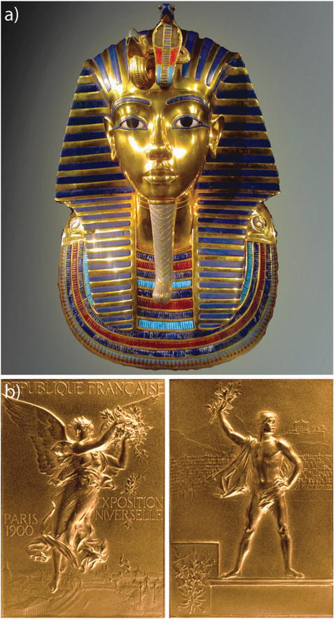
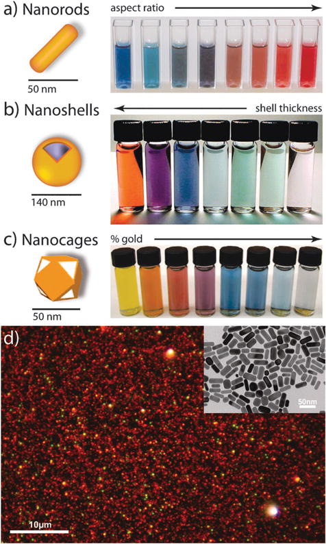
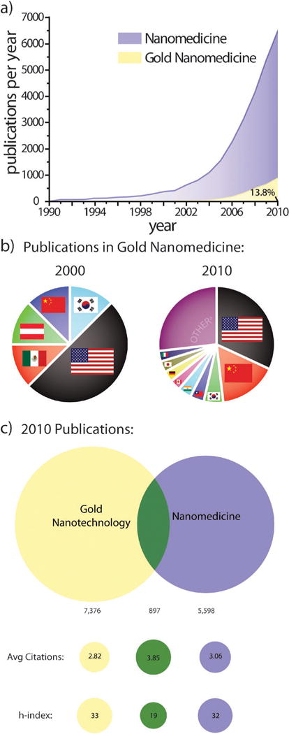
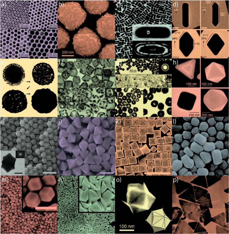
Gold nanoparticles have been used in biomedical applications since their first colloidal syntheses more than three centuries ago. However, over the past two decades, their beautiful colors and unique electronic properties have also attracted tremendous attention due to their historical applications in art and ancient medicine and current applications in enhanced optoelectronics and photovoltaics. In spite of their modest alchemical beginnings, gold nanoparticles exhibit physical properties that are truly different from both small molecules and bulk materials, as well as from other nanoscale particles. Their unique combination of properties is just beginning to be fully realized in range of medical diagnostic and therapeutic applications. This critical review will provide insights into the design, synthesis, functionalization, and applications of these artificial molecules in biomedicine and discuss their tailored interactions with biological systems to achieve improved patient health. Further, we provide a survey of the rapidly expanding body of literature on this topic and argue that gold nanotechnology-enabled biomedicine is not simply an act of 'gilding the (nanomedicinal) lily', but that a new 'Golden Age' of biomedical nanotechnology is truly upon us. Moving forward, the most challenging nanoscience ahead of us will be to find new chemical and physical methods of functionalizing gold nanoparticles with compounds that can promote efficient binding, clearance, and biocompatibility and to assess their safety to other biological systems and their long-term term effects on human health and reproduction (472 references).


Figures

Fig. 1
Fig. 1
(a) Golden burial mask of Egyptian Pharaoh Tutankhamun (King Tut) of the 18th Dynasty (ca. 1323 BC). (b) A gold medal presented at the Games of the II Olympiad (Paris, France; 1900). While bulk gold is highly un-reactive and predominantly reflects light, nanoscale gold can be highly reactive, exhibiting pharmacologic properties and the ability to absorb, transfer, and convert light energy into heat. The mask in (a), discovered in 1922 by Howard Cater, consists of solid gold with inlaid glass and stone (21 cm high and ca. 11 kg). Prior to the 1900 Olympics in (b), athletes received only silver and copper medals which easily oxidize. The winged goddess Nike is shown on the front in (b); a victorious athlete holding a laurel branch is shown on the back with The Acropolis in the background. Image (a) by James A. Buckley. Image (b) reprinted with permission from the International Olympic Committee. Copyright IOC.
Fig. 2
Fig. 2
Gold nanoparticles commonly applied in biomedical applications. (a) Gold nanorods, (b) silica–gold core–shell nanoparticles, and (c) gold nanocages. The intense color of these nanoparticles arises from the collective excitation of their conduction electrons, or surface plasmon resonance modes, which results in photon absorption at wavelengths which varies with (a) aspect ratio, (b) shell thickness, and/or (c) galvanic displacement by gold. (d) Optical dark-field scattering micrograph of gold nanorods (electron micrograph in the inset) showing resonant scattering from their transverse (short-axis) plasmon mode (green) and their lower energy, longitudinal (long-axis) plasmon mode (red)). Image (a) by X. Huang, (b) by C. Radloff and N.J. Halas, and (d) by C. Rosman and C. Sönnichsen. Figures adapted with permission from (b) ref. and (c) ref. . Copyright (a) 2003 Annual Reviews and (b) 2007 Macmillan Publishers Ltd.: Nature Publishing Group.
Fig. 3
Fig. 3
Worth more than its weight: exponential growth in the number of publications on gold nanotechnology and nanomedicine over the past two decades. (a) Annual publications in nanomedicine dramatically increased following award of the 1996 Nobel Prize in Chemistry to Kroto, Curl, and Smalley for their discovery of fullerenes. Medicinal applications of gold nanotechnologies further added to this growth following US President Bill Clinton’s formation of the National Nanotechnology Infrastructure Network (NNIN) in 2000 and US President George H. W. Bush’s expansion of the program in 2003 with the 21st Century Nanotechnology Research and Development Act. (b) Contributions from various countries to publications on gold nanomedicine in 2000 and 2010. Publications in 2000 were limited to just 5 countries while those in 2010 included more than 50. Other countriesa represent those with <2.9%. (c) Overlap between publications on gold nanotechnology and nanomedicine in 2010 and comparison of their corresponding average number of citations and h-indices. Note that publication data in (a) is not cumulative.
Fig. 4
Fig. 4
Gold nanoparticles of various size and shape with potential applications in biomedicine. Small (a) and large (b) nanospheres, (c) nanorods, (d) sharpened nanorods, (e) nanoshells, (f) nanocages/frames, (g) hollow nanospheres, (h) tetrahedra/octahedra/cubes/icosahedra, (i) rhombic dodecahedra, (j) octahedra, (k) concave nanocubes, (l) tetrahexahedra, (m) rhombic dodecahedra, (n) obtuse triangular bipyramids, (o) trisoctahedra, and (p) nanoprisms. Figures adapted with permission from (a) ref. , (b) ref. , (c) ref. and , (d) ref. , (e) ref. , (f) ref. , (g) ref. , (h) ref. , (i–j) ref. , (k) ref. , (l) ref. , (m–n) ref. , (o) ref. , and (p) ref. . Copyright (a) 2003 American Chemical Society, (b) 2008 Wiley-VCH Verlag GmbH & Co., (c) 2004 American Chemical Society and 1999 Elsevier Science B.V., (d) 2007 Wiley-VCH Verlag GmbH & Co., (e) 1998 Elsevier Science B.V., (f) 2007 American Chemical Society, (g) 2005 American Chemical Society, (h) 2004 Wiley-VCH Verlag GmbH & Co., (i–j) 2009 American Chemical Society, (k) 2010 American Chemical Society, (l) 2009 American Chemical Society, (m–n) 2011 American Chemical Society, (o) 2008 VCH Verlag GmbH & Co., and (p) 2005 American Chemical Society.
Fig. 5
Fig. 5
Exemplary gold nanostructures obtained by various “top-down” synthetic approaches. (a) Nanosphere lithography (NSL), (b) electron-beam lithography (EBL), (c–e) nanoskiving, (f–i) dip-pen lithography (DPL), (j–l) structural transformation by electro-deposition on patterned substrates (STEPS), (m–o) nanocrescent synthesis, and (p–s) nanopyramid synthesis. Figures adapted with permission from (a) ref. , (b) ref. , (c–e) ref. , (f–i) ref. , (j–l) ref. , (m–o) ref. , and (p–s) ref. , . Copyright (a) 2005 American Chemical Society, (b) 2011 American Institute of Physics, (c–e) 2008 American Chemical Society, (f–i) 2004 American Chemical Society, (j–l) 2011 American Chemical Society, (m–o) 2005 American Chemical Society, (p) 2007 American Chemical Society, and (q–s) 2008 American Chemical Society.
Fig. 6
Fig. 6
Comparison of dithiolate and thiolate oxidative desorption from gold nanoparticles (a) over a 73 hour period (b). Figure/data adapted with permission from ref. . Copyright 2009 American Chemical Society.
Fig. 7
Fig. 7
Schematics illustrating various methods by which gold nanoparticles can be conjugated with biofunctional molecules. (a) hydrophobic entrapment, (b) electrostatic adsorption, and (c) covalent cross coupling by carbodiimide, maleimide, and click chemistry. Figures adapted with permission from (a) ref. and (c) ref. , , and . Copyright (a) 2009 American Chemical Society and (c) 2007 American Chemical Society and 2010 Wiley-VCH Verlag GmbH & Co.
Fig. 8
Fig. 8
Schematics illustrating additional methods by which gold nanoparticles can be conjugated with biofunctional molecules. (a) dative covalent bonding, (b) oligonucleotide hybridization, and (c) and photolabile linkage. Figures adapted with permission from (a) ref. , , (b) ref. , and (c) ref. . Copyright (a) 2010 and 2009, (b) 2007, and (c) 2009 American Chemical Society.
Fig. 9
Fig. 9
Silane conjugation chemistry for biomedical gold nanoparticle conjugates. Silica shell (Stöber) functionalized (a) gold nanospheres and (b) gold nanoprisms. Reaction schemes (c) for conjugation to (i) hydroxyl-and (ii) silane-functionalized gold nanoparticles. Reaction scheme (d) for the encapsulation of bioanalytically- and/or therapeutically-relevant molecules about gold nanoparticles. Figures adapted with permission from (a) ref. (b) ref. (c) ref. and (d) ref. . Copyright (a) 1996, (b) 2010, (c) 2007, and (d) 2003 American Chemical Society.
Fig. 10
Fig. 10
SERS detection of cancer cells using immunolabeled gold nanorods. (a) SERS spectra of normal HaCaT cells incubated with anti-EGFR antibody conjugated gold nanorods. (b) SERS spectra of HSC cancer cells incubated with anti-EGFR antibody conjugated gold nanorods. Cancer cells in (b) show stronger, sharper and better resolved SERS signals than normal cells in (a) due to the specific binding of immunolabeled gold nanorods with receptors on the cancer cell surface, suggesting that SERS may serve as a clinical diagnostic tool. The sharper and stronger Raman signals in (b) result from electromagnetic field enhancement due to interparticle coupling between immunolabeled nanorods and their alignment along the cellular membrane surface. Figures adapted with permission from ref. . Copyright 2007 American Chemical Society.
Fig. 11
Fig. 11
SERS detection of circulating tumor cells in patient blood samples. (a) Schematic illustration of SERS nanoparticles and their conjugation with epidermal growth factor peptides. (b) SERS spectra of different numbers of Tu212 cancer cells spiked into mouse white blood cells. (c) SERS spectra of blood sample from a patient incubated with targeted and non-targeted SERS nanoparticles, as well as a blood sample from a healthy donor incubated with targeted SERS nanoparticles. The SERS nanoparticles can detect circulating tumor cells with a sensitivity of 5–50 cells per mL blood. The strong signals from cancer patient indicates highly specific and sensitive detection of circulating tumor cells in blood system. Figures adapted with permission from ref. . Copyright 2011 American Association for Cancer Research.
Fig. 12
Fig. 12
A single particle localized surface plasmon resonance (LSPR) assay for biomolecular detection. (a) Electron micrographs of gold nanorods. (b) Optical dark-field scattering microscopy of gold nanorods. (c) Real-time measurement of the binding of streptavidin and biotin by monitoring the light scattering spectra of a single biotin-conjugated gold nanorod. Black, blue, and red curves represent the spectral shift over time when biotin-conjugated gold nanorods were incubated in 130 nM, 10 nM and 1 nM streptavidine in PBS. The gold nanorods showed a sensitivity of streptavidin detection down to 1 nM. Figures adapted with permission from ref. . Copyright 2008 American Chemical Society.
Fig. 13
Fig. 13
DNA detection using gold nanoparticles. (a) Schematic illustration of DNA detection based on hybridization-induced gold nanoparticle aggregation. (b) Visualization of gold nanoprobes with and without the presence of target DNA. (c) Monitoring the aggregation process by spotting the solution on a silica support. Black and red curves: in the presence of complementary DNA target, the oligonucleotides on the surfaces of the gold nanoparticles will bind to the target and induce aggregation of gold nanoparticles and a blue color change. Figures adapted with permission from ref. and . Copyright 1997 American Association for the Advancement of Science and 2005 American Chemical Society.
Fig. 14
Fig. 14
Cancer diagnostics using gold nanorod-enhanced light scattering. Optical dark-field microscopy of normal HaCaT cells and cancerous HSC and HOC cells incubated with anti-EGFR antibody-conjugated gold nanospheres (top panels, left to right). Optical dark-field microscopy of normal HaCaT cells and cancerous HSC and HOC cells incubated with anti-EGFR antibody-conjugated gold nanorods (lower panel, left to right). Anti-EGFR conjugated gold nanoparticles specifically bound to cancer cells, scattering strongly under dark-field microscopy and thus enabling detection of malignant cells. Figures adapted with permission from ref. . Copyright 2006 American Chemical Society.
Fig. 15
Fig. 15
Tumor diagnosis by using gold nanoshell-enhanced optical coherence tomography imaging. Optical coherent tomography image of (a) normal tissue injected with saline, (b) normal tissue injected with gold nanoshells, (c) tumor tissue injected with saline, and (d) tumor tissue injected with gold nanoshells. A significant increase in image contrast from tumor tissue is observed compared with normal tissue or tumor tissue injected with PBS solution. Figures adapted with permission from ref. . Copyright 2007 American Chemical Society.
Fig. 16
Fig. 16
Two-photon luminescence (TPL) imaging of the accumulation and uptake of small-molecule targeted gold nanorods. (A) KB cancer cells incubated with folate-conjugated gold nanorods for 6 h. (B) KB cancer cells incubated with folate-conjugated gold nanorods for 17 h. (C) Normal NIH-3T3 cells incubated with folate-conjugated gold nanorods. Due to the strong two photon luminescence signals from gold nanorods, the location of the nanorods could be clearly visualized. At 6 h, the nanorods were accumulated on the cell membrane of cancer cells. At 17 h, the nanorods were internalized into the cancer cells. Functionalized gold nanorods did not show nonspecific binding to normal cells. Figures adapted with permission from ref. . Copyright 2007 Wiley-VCH Verlag GmbH & Co.
Fig. 17
Fig. 17
Photoacoustic imaging of blood vessels in the mouse brain using gold nanocages. (A) Photoacoustic image of a mouse brain larger (yellow-framed picture) and small (green-framed picture) blood vessels 2 h after intravenous injection of poly(ethylene glycol)-conjugated gold nanocages. (B–D) Optical images of mouse brain vessels 2 h after injecting poly(ethylene glycol)-conjugated gold nanocages. Blood vessels are stained with anti-CD13 antibody (red). Gold nanocages are imaged by dark-field scattering microscopy (pseudogreen). Gold nanocages enhanced the photoacoustic signals of blood vessels in the mouse brain, revealing a clear and detailed structure vasculature as small as 100 μm in diameter. Figures adapted with permission from ref. . Copyright 2010 Elsevier B.V.
Fig. 18
Fig. 18
Laser photothermal cancer therapy using gold nanorod contrast agents. PEGylated gold nanorods were intravenously and locally injected in carcinoma-bearing mice. (a) NIR transmission images obtained with a simple cell phone camera and an inexpensive NIR-diode laser show substantial laser attenuation due to absorption by nanorods accumulated at the rear flank tumor site. (b) Change in tumor volume over two weeks following a single laser exposure, indicating significant tumor growth remission for both direct and intravenous nanorod administration, as well as resorption of >57% of the directly-injected tumors and 25% of the intravenously-treated tumors. (c) Real-time, intratumoral thermal transient measurements correlating enhanced heating of nanorod-treated tumors with tumor resorption/remission. Reprinted with permission from ref. . Copyright 2008 Elsevier Science B.V.
Fig. 19
Fig. 19
Illustration demonstrating various approaches to loading/ unloading therapeutics into/from gold nanoparticles. Partitioning and diffusion-driven release of hydrophobic drug molecules in (a) a surfactant bilayer or (b) an amphiphilic corona layer. (c) Anchoring drugs directly to the surfaces of gold nanoparticles through Au–S or Au–N bonds (capping agent in blue is hydrophilic polymer, e.g. PEG, to enhance the overall solubility of the system). Release is triggered by the photothermal effect, thiol exchange (e.g. glutathione exchange), or simple diffusion to the cell membranes (in the case of Au–N). (d–e) Double-stranded DNA-loaded gold nanoparticles via Au–S bonding. The release of double (d) or single (e) stranded DNA is controlled by an applied laser. (f) Therapeutic agents are coupled/complexed to terminal functional groups of the capping agent via a cleavable linker. Release can be triggered by hydrolysis, light, heat, and/or pH changes. (g) Loading charged biomolecules (e.g. DNA or siRNA) onto the surfaces of gold nanoparticles by electrostatic assembly (LbL coating, see text for details). Release of payload can be triggered by the use of charge-reversal polyelectrolytes combined with pH change. (h) Drug molecules are incorporated into the matrix of a thermosensitive, crosslinked polymer. Release can be triggered by the photothermal heating by gold nanoparticles also incorporated into the matrix.
Fig. 20
Fig. 20
Enhanced breast cancer drug delivery using tamoxifen-gold nanoparticle conjugates. (a) A thiol-PEGyalted derivative of the estrogen receptor antagonist, tamoxifen, was conjugated to gold nanoparticles, allowing (b) increasingly rapid and selective drug delivery to breast cancer cells which overexpress the hormone receptor, estrogen receptor. (c) Dark-field scattering microscopy of breast cancer cells showing estrogen receptor-selective intracellular particle delivery. (d–e) Dose–response kinetics indicate accelerated drug transport rates via nanoparticle endocytosis versus passive diffusion of the free drug, resulting in >104-fold enhanced potency (2.7-fold per drug molecule). Reprinted with permission from ref. Copyright 2009 American Chemical Society.
Fig. 21
Fig. 21
Examples of loading drugs into interior reservoirs of gold nanoparticles. (a) Gold nanocages (hollow gold cubes with porous walls) are functionalized with a thermosensitive polymer brush layer at their exterior surface to cage drug molecules in their interior. Laser irradiation induces local heat flux and thus, collapse of the thermo-sensitive polymer to release the caged drug molecules. (b) Gold nanocages with the drugs dispersed into a thermosensitive material in the interior of the nanoparticles. Laser irradiation results in phase-change (melting) of the thermosensitive “filler” and thus enhances drug release. (c) A gold nanoshell covers a liposome carrying drugs in its interior. Gold nanoshells absorb light and convert it to heat and these events result in disintegration and clearance of the carrier, as well as release of its encapsulated drugs.
Fig. 22
Fig. 22
(a) Illustration demonstrating the use of gold nanoparticles in a composite material to enable light-triggered drug delivery. Gold nanorods distributed in a polymeric microsphere matrix act as localized nanoheaters upon light irradiation. Gold nanorods absorb light and convert it into heat which changes the polymeric matrix from a glassy state to a rubbery state and allowing enhanced drug diffusion and release. (b) Experiential results showing drug release as a function of laser irradiation cycles/duration for a microsphere matrix containing gold nanorods. Squares: with laser; X: no laser. Laser λmax = 808 nm; Tg = glass transition temperature. Panel (b) is adapted with permission from ref. . Copyright 2011 American Chemical Society.
Fig. 23
Fig. 23
Illustration demonstrating potential sources of data artifacts obtained when performing in vitro cellular toxicity and uptake studies with nanomaterials. (a) Toxicity could be due to free chemicals in solution and not to the particles themselves; thus, comparing the toxicity of nanoparticle solution with its supernatant is an important control. (b) Nanoparticles could adsorb to the cell surface (on cells) or enter to the inside (in cells). Quantification of gold content in collected cells cannot differentiate between both types of interactions and may result in an overestimated uptake. (c) Differential cellular uptake of nanoparticles could be due to different sedimentation rates. Nanoparticles with a high sedimentation rate (c, left) reach the nanoparticle– cell interaction zone faster than nanoparticles with a low sedimentation rate (c, right) and thus exhibit higher uptake. Ignoring this factor could result in erroneous correlation between uptake and other factors such as size, charge, and surface chemistry.
Fig. 24
Fig. 24
(a) Illustration demonstrating binding of gold nanoparticles (G2, G40, G70 for 2, 40, 70 nm diameter) functionalized with Herceptin antibodies, which recognize receptors on the cell surface (HER2/neu, ErbB2). G40 interacts with the receptors more efficiently due to its unique size and propensity for endocytosis. Lower panel: fluorescence images of the cellular distribution of ErbB2 (red) after treatment with fluorescently-labeled G2, G40, and G70. Note that only in the case of G40 treatment, that particles redistributed form the cell surface to the cytoplasm due to efficient endocytosis. (b) Increase in relative fluorescence intensity following nanoparticle uptake was found to correlate with (c) subsequent cell death. Nuclei are stained blue, scale bar = 10 mm, *p < 0.05, error bars ±sd, n = 4. Adapted with permission from ref. . Copyright 2008 Macmillan Publishers Ltd.: Nature Publishing Group.
Fig. 25
Fig. 25
Illustration of a biodegradable nanoparticle–polymer composite. Gold nanoparticles (4 nm in diameter) are used as building blocks to form NIR-absorbing plasmonic nanoparticle upon interaction with a biodegradable polymer. The formed nanoclusters can be dissociated to smaller aggregates and ultimately to their initial building blocks by pH drop inside acidic compartments of the cell. Disintegration of nanoparticles to smaller fragments is advantageous to enhance the total urinary clearance of nanoparticles from the body. Adapted with permission from ref. . Copyright 2010 American Chemical Society.

References

    1. Greenwood NN, Earnshaw A. Chemistry of the Elements. 2nd. Butterworth-Heinemann; Burlington, MA: 1997.
    1. Chen MS, Goodman DW. Science. 2004;306:252–255. - PubMed
    1. Valden M, Lai X, Goodman DW. Science. 1998;281:1647–1650. - PubMed
    1. Green IX, Tang W, Neurock M, Yates JT. Science. 2011;333:736–739. - PubMed
    1. Shaw CF. Chem Rev. 1999;99:2589–2600. - PubMed

Publication types