Comparative Study
. 2012 May;55(5):1529-39.
doi: 10.1002/hep.24815. Epub 2012 Apr 4.

The microenvironment in hepatocyte regeneration and function in rats with advanced cirrhosis

Affiliations
Comparative Study

The microenvironment in hepatocyte regeneration and function in rats with advanced cirrhosis

Liping Liu et al. Hepatology. 2012 May.

Abstract

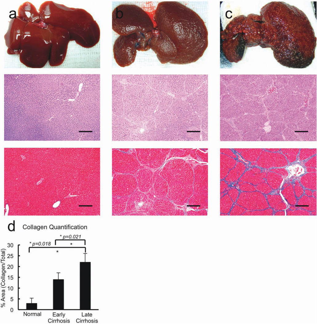
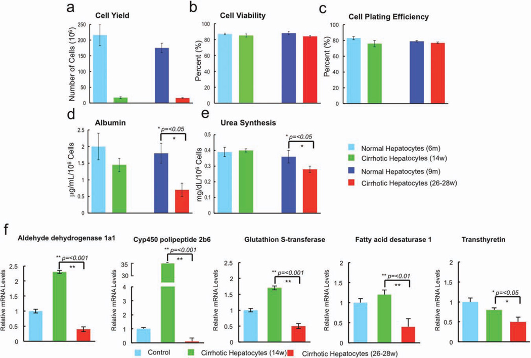
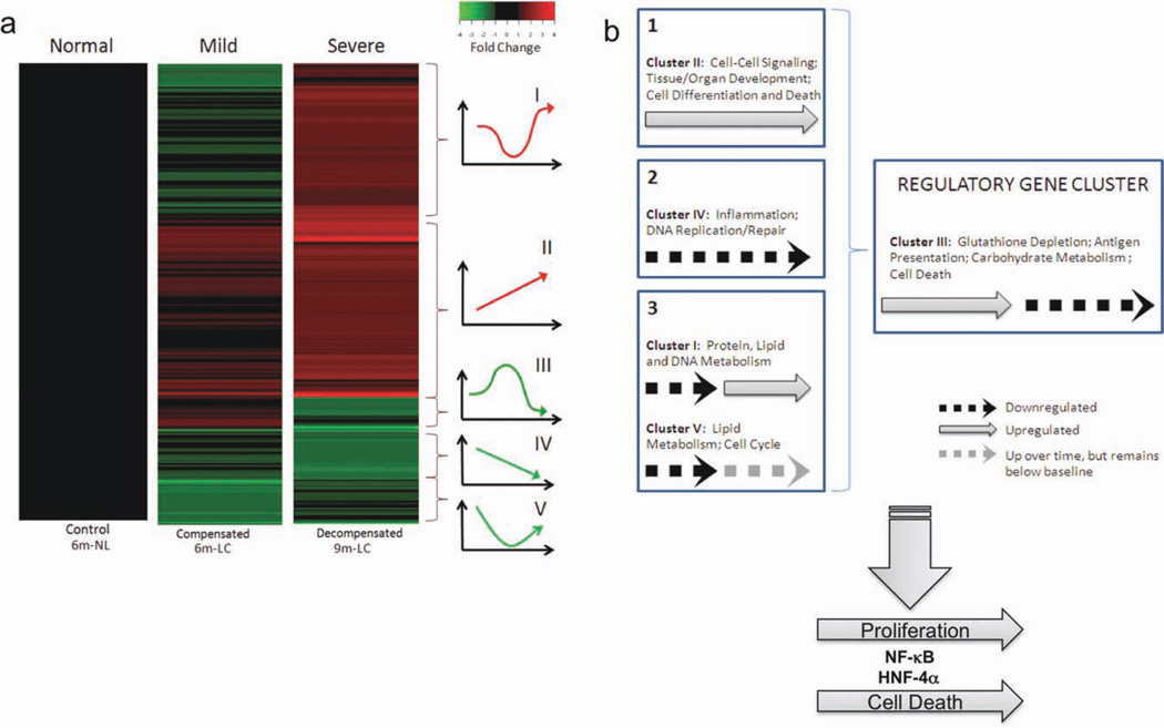
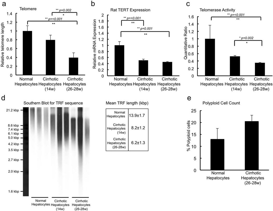
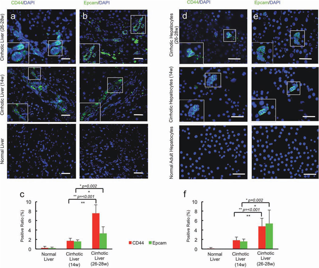
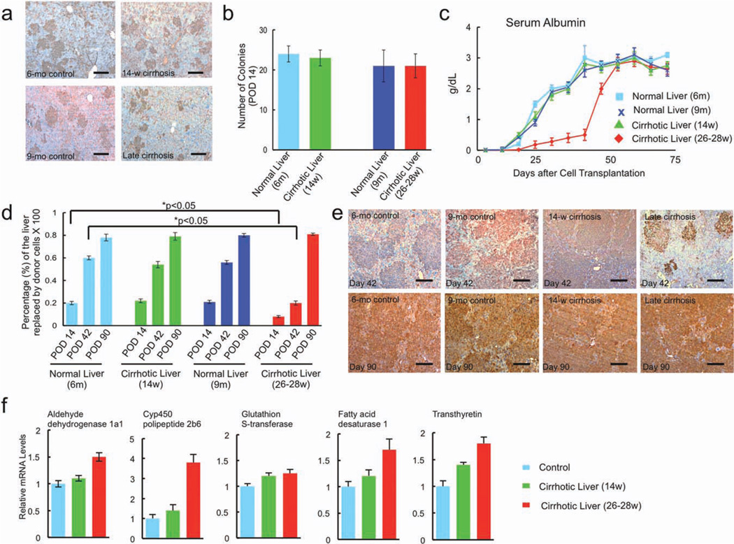
In advanced cirrhosis, impaired function is caused by intrinsic damage to the native liver cells and from the abnormal microenvironment in which the cells reside. The extent to which each plays a role in liver failure and regeneration is unknown. To examine this issue, hepatocytes from cirrhotic and age-matched control rats were isolated, characterized, and transplanted into the livers of noncirrhotic hosts whose livers permit extensive repopulation with donor cells. Primary hepatocytes derived from livers with advanced cirrhosis and compensated function maintained metabolic activity and the ability to secrete liver-specific proteins, whereas hepatocytes derived from cirrhotic livers with decompensated function failed to maintain metabolic or secretory activity. Telomere studies and transcriptomic analysis of hepatocytes recovered from progressively worsening cirrhotic livers suggest that hepatocytes from irreversibly failing livers show signs of replicative senescence and express genes that simultaneously drive both proliferation and apoptosis, with a later effect on metabolism, all under the control of a central cluster of regulatory genes, including nuclear factor κB and hepatocyte nuclear factor 4α. Cells from cirrhotic and control livers engrafted equally well, but those from animals with cirrhosis and failing livers showed little initial evidence of proliferative capacity or function. Both, however, recovered more than 2 months after transplantation, indicating that either mature hepatocytes or a subpopulation of adult stem cells are capable of full recovery in severe cirrhosis.

Conclusion: Transplantation studies indicate that the state of the host microenvironment is critical to the regenerative potential of hepatocytes, and that a change in the extracellular matrix can lead to regeneration and restoration of function by cells derived from livers with end-stage organ failure.


Conflict of interest statement

Potential conflict of interest: Dr. Kaestner consults for Johnson & Johnson.

Figures

Fig. 1
Fig. 1
Gross and histologic changes associated with CCl4-induced liver injury. (a) Examination of normal control livers, (b) early cirrhotic livers from animals treated with 14 weeks of CCl4, and (c) advanced cirrhotic livers from animals treated with 26–28 weeks of CCl4 were examined via gross examination (upper panels), hematoxylin and eosin (middle panels; bar = 250 µm), and Masson’s trichrome stain (lower panels; bar = 250 µm). Cirrhotic livers contained numerous regenerating nodules on gross inspection. Histologic analysis documented nodular regenerative hyperplasia and cirrhosis in both groups of animals although fibrosis and (d) collagen deposition was more extensive in animals that received the greater amount of CCL4.
Fig. 2
Fig. 2
Yield after isolation and functional characteristics of cells recovered from normal control livers, early cirrhotic livers, and advanced cir-rhotic livers. (a) The yield of cells recovered by collagenase digestion from cirrhotic livers was significantly lower than that recovered from age-matched controls and was approximately 5% of that recovered from control livers. (b,c) Cell viability (b) and cell plating efficiency (c) were not statistically different among groups. (d,e) Hepatocytes derived from control rats and rats with compensated cirrhosis secreted equal amounts of albumin (d) and urea (e), whereas hepatocytes from the livers of cirrhotic rats with liver failure secreted significantly less of each (P < 0.05). (f) A cohort of liver-specific genes was examined using qPCR and documented up-regulation in early cirrhosis followed by significant down-regulation (compared with control) in late cirrhosis of CYP450 and metabolic enzyme gene expression in hepatocytes derived from the livers of rats with decompensated cirrhosis: ADH1a1, CYP4502b9, GST, FADS1, and transthyretin.
Fig. 3
Fig. 3
DNA microarray analysis of messenger RNA from hepatocytes recovered from normal control, early cirrhotic, and advanced cirrhotic livers. (a) Hierarchical clustering of differentially expressed genes from hepatocytes isolated from livers with compensated and decompensated cirrhosis demonstrated significant gene expression differences among groups depending on the extent of cirrhosis from which the hepatocytes were derived. (b) Schematic representing hypotheses regarding liver cirrhosis, as gleaned from gene expression data. The DNA microarray results suggest that the irreversibly cirrhotic liver is expressing genes that simultaneously drive both proliferation and apoptosis, with a later effect on metabolism (clusters I, II, IV, and V ) , under the control of a central cluster of regulatory genes (cluster III). This regulatory process involves the actions of NF-κB and HNF-4α.
Fig. 4
Fig. 4
Assessment of markers for senescence in hepatocytes recovered from normal control, early cirrhotic, and advanced cirrhotic livers. (a,b) qPCR for telomere length (a) and rat telomerase expression (b) showed progressive loss of activity with progression of cirrhosis. These data were confirmed by direct assessment of functional telomerase activity (c), and Southern blot analysis (d) demonstrated critical shortening of telomere length in hepatocytes derived from end-stage cirrhotic livers. These studies indicate the occurrence of replicative senescence, because cirrhosis leads to decompensated liver function. (e) An increase in polyploidy was also noted.
Fig. 5
Fig. 5
Induction of hepatic progenitor cells in progressively worsening cirrhosis. (a–c) As cirrhosis progressed, there was an associated increase in the percentage of cells expressing putative liver progenitor cell markers (a) CD44 and (b) Epcam in liver sections bar = 100 µm. **P ≤ 0.05. Hepatocytes isolated from control, early cirrhotic, and end-stage cirrhotic livers, which were used in transplantation studies, were similarly characterized for the presence of putative liver progenitor cells. (d–f) A nearly identical percentage of the cells isolated from cirrhotic livers expressed CD44 (d) and Epcam bar = 100 µm (e) in liver sections. These data indicate that the distribution of cell phenotypes derived from cirrhotic livers after isolation most likely represented that found in intact livers even though the cell yield after collagenase digestion was significantly lower than that obtained after digestion of control livers.
Fig. 6
Fig. 6
Repopulation of retrorsine-treated Nagase analbuminemic rat livers by donor hepatocytes. (a) Fourteen days after transplantation, donor cells from age-matched control, early cirrhotic, and advanced cirrhotic livers appeared to engraft with equal capacity bar = 150 µm. The albumin-expressing hepatocyte colonies were relatively small in size, as expected, and their numbers (b) were not significantly different among groups. (c) Early after transplantation, serum albumin levels in rats that received cells derived from donors with early cirrhosis and controls were significantly higher than in rats that received cells from cirrhotic rats with liver failure. The serum albumin levels in recipients with cell transplants from failing cirrhotic livers, however, recovered their capacity to expand and release albumin approximately 2 months after engraftment in noncir-rhotic livers. (d) Fourteen days after transplantation, there was a small difference in the percentage of the liver replaced by donor hepatocytes from control and early cirrhotic livers compared with that replaced by donor hepatocytes from failing cirrhotic livers (P < 0.05). (e) By posttransplantation day 42, there was considerable expansion of transplanted hepatocytes derived from controls and livers with early cirrhosis with coalescence of hepatocyte colonies, whereas expansion of transplanted hepatocytes recovered from failing cirrhotic livers was significantly less. By posttransplantation day 90, however, approximately 80% of the liver of all recipient rats was replaced by albumin-producing hepatocytes, independent of the source of the donor cells. Expression levels of previously measured liver-specific genes were assessed to determine whether recovered donor hepatocytes normalize function after transplantation. (f) qPCR demonstrated essentially no difference in ADH1a1, CYP4502b9, GST, FADS1, and transthyretin in the livers of repopulated animals irrespective of the source of donor cells used for transplantation.

References

    1. Iredale JP, Benyon RC, Pickering J, McCullen M, Northrop M, Pawley S, et al. Mechanisms of spontaneous resolution of rat liver fibrosis. Hepatic stellate cell apoptosis and reduced hepatic expression of metalloproteinase inhibitors. J Clin Invest. 1998;102:538–549. - PMC - PubMed
    1. Zeisberg M, Kalluri R. Reversal of experimental renal fibrosis by BMP7 provides insights into novel therapeutic strategies for chronic kidney disease. Pediatr Nephrol. 2008;23:1395–1398. - PubMed
    1. Teixeira-Clerc F, Julien B, Grenard P, Tran Van Nhieu J, Deveaux V, Li L, et al. CB1 cannabinoid receptor antagonism: a new strategy for the treatment of liver fibrosis. Nat Med. 2006;12:671–676. - PubMed
    1. Du XJ, Xu Q, Lekgabe E, Gao XM, Kiriazis H, Moore XL, et al. Reversal of cardiac fibrosis and related dysfunction by relaxin. Ann N Y Acad Sci. 2009;1160:278–284. - PubMed
    1. Schuppan D, Afdhal NH. Liver cirrhosis. Lancet. 2008;371:838–851. - PMC - PubMed

Publication types

MeSH terms