. 2013 Apr;34(4):837-51.
doi: 10.1002/hbm.21478. Epub 2011 Nov 23.

Cortical oscillatory changes in human middle temporal cortex underlying smooth pursuit eye movements

Affiliations

Cortical oscillatory changes in human middle temporal cortex underlying smooth pursuit eye movements

Benjamin T Dunkley et al. Hum Brain Mapp. 2013 Apr.

Abstract

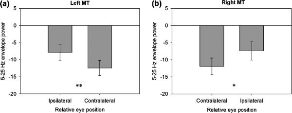
Extra-striate regions are thought to receive non-retinal signals from the pursuit system to maintain perceptual stability during eye movements. Here, we used magnetoencephalography (MEG) to study changes in oscillatory power related to smooth pursuit in extra-striate visual areas under three conditions: 'pursuit' of a small target, 'retinal motion' of a large background and 'pursuit + retinal motion' combined. All stimuli moved sinusoidally. MEG source reconstruction was performed using synthetic aperture magnetometry. Broadband alpha-beta suppression (5-25 Hz) was observed over bilateral extra-striate cortex (consistent with middle temporal cortex (MT+)) during all conditions. A functional magnetic resonance imaging study using the same experimental protocols confirmed an MT+ localisation of this extra-striate response. The alpha-beta envelope power in the 'pursuit' condition showed a hemifield-dependent eye-position signal, such that the global minimum in the alpha-beta suppression recorded in extra-striate cortex was greatest when the eyes were at maximum contralateral eccentricity. The 'retinal motion' condition produced sustained alpha-beta power decreases for the duration of stimulus motion, while the 'pursuit + retinal motion' condition revealed a double-dip 'W' shaped alpha-beta envelope profile with the peak suppression contiguous with eye position when at opposing maximum eccentricity. These results suggest that MT+ receives retinal as well as extra-retinal signals from the pursuit system as part of the process that enables the visual system to compensate for retinal motion during eye movement. We speculate that the suppression of the alpha-beta rhythm reflects either the integration of an eye position-dependent signal or one that lags the peak velocity of the sinusoidally moving target.


Figures

Figure 1
Figure 1
Schematic of experimental protocol for the MEG experiment. fMRI parameters are detailed within the text. (a) ‘Pursuit’: a single stationary low‐contrast, monochromatic dot was presented as a fixation point for the 10‐s passive/rest phase. The dot then oscillated back and forth sinusoidally in the horizontal plane for 10 s at ±5° at a frequency of 0.5 Hz, followed by another passive period consisting of 10‐s fixation. This was repeated for 30 trials. (b) ‘Retinal motion’ condition: a central dot was fixated for 10 s (rest), followed by 10 s of an oscillating background consisting of a random dot field. (c) ‘Pursuit + retinal motion’: fixation was maintained for 10 s, followed by pursuit over a stationary random dot field (note that window aperture moved in the same way as the pursuit target).
Figure 2
Figure 2
Mean eye velocity gain (ratio of eye velocity to stimulus velocity) for all three conditions.
Figure 3
Figure 3
Group results from MEG data and fMRI data in all three conditions, overlaid on a template brain. (a) MEG data, left panel: group‐averaged SAM images in the 15–25 Hz frequency band during smooth pursuit eye movements, overlaid on a template brain. Oscillatory peaks occur in bilateral extra‐striate cortex, with notable beta suppression in areas consistent with putative MT+. Blue/purple/white colour map indicates negative oscillatory amplitude changes with colour bar showing pseudo‐t values. fMRI data, right panel: group fMRI data showing significantly activated clusters (P < 0.05) during pursuit, with warm colours indicating an increasing BOLD amplitude. Note the similar spatial locations of the beta suppression in the MEG data and the BOLD effect in the fMRI data. (b) Group SAM images (15–25 Hz) during ‘retinal motion’ (condition 2) and fMRI data. (c) Group SAM images for ‘pursuit + retinal motion’ (condition 3) and fMRI data. [Color figure can be viewed in the online issue, which is available at wileyonlinelibrary.com.]
Figure 4
Figure 4
(a) Grand‐averaged time–frequency spectrograms during the ‘pursuit’ condition, extracted from the extra‐striate virtual sensor location in both left and right hemispheres, showing task‐induced broadband alpha–beta activity decreases for the duration of target tracking. (b) Spectrograms for ‘retinal motion’ and (c) ‘pursuit + retinal motion’ conditions. [Color figure can be viewed in the online issue, which is available at wileyonlinelibrary.com.]
Figure 5
Figure 5
(a) Horizontal eye position data for the duration of object tracking and single pursuit cycle average eye position. (b) Group‐averaged 5–25 Hz envelope oscillatory amplitude change during pursuit from the left hemisphere MT+ voxel, with single pursuit cycle alpha–beta amplitude average. Maximum suppression of the rhythm appears to correspond to pursuit position when the eye gaze was at maximum eccentricity in the contralateral visual hemifield. (c) Same as (b), except the 5–25 Hz amplitude envelope from right hemisphere MT+ voxel. [Color figure can be viewed in the online issue, which is available at wileyonlinelibrary.com.]
Figure 6
Figure 6
(a) Average broadband alpha–beta amplitude from left MT+ during smooth pursuit, quantified over a 100‐ms window when eyes were at maximum amplitude, relative to virtual electrode location. (b) Same as before, for right MT+. **P < 0.001, *P < 0.05.
Figure 7
Figure 7
(a) Stimulus position (moving dot‐field pattern; red trace) during retinal motion and eye position (blue trace) and single stimulus position cycle with average eye position. (b) Group‐averaged 5–25 Hz amplitude envelope change during retinal motion from the left hemisphere MT+ voxel, with single stimulus cycle alpha–beta amplitude average. Alpha–beta rhythm suppression appears largely sustained for the duration of stimulus motion. (c) Same as (b), except 5–25 Hz amplitude envelope from right hemisphere MT+ voxel. [Color figure can be viewed in the online issue, which is available at wileyonlinelibrary.com.]
Figure 8
Figure 8
(a) Eye position during pursuit and single cycle eye position average. (b) Group‐averaged 5–25 Hz amplitude envelope change during pursuit over a stationary background from the left hemisphere MT+ voxel, with single stimulus cycle alpha–beta amplitude average. Peak alpha–beta activity decrease occurs with maximum eye eccentricity in the contraleral visual hemifield, with a second peak decrease appearing to reflect eye position in the ipsilateral hemifield during pursuit. (c) Same as (b), except 5–25 Hz amplitude envelope from right hemisphere MT+ voxel. [Color figure can be viewed in the online issue, which is available at wileyonlinelibrary.com.]

References

    1. Baillet S, Mosher JC, Leahy RM ( 2001): Electromagnetic brain mapping. IEEE Signal Process Mag 18: 14–30.
    1. Barnes GR, Hillebrand A ( 2003): Statistical flattening of MEG beamformer images. Hum Brain Mapp 18: 1–12. - PMC - PubMed
    1. Brookes MJ, Gibson AM, Hall SD, Furlong PL, Barnes GR, Hillebrand A, Singh KD, Holliday IE, Francis ST, Morris PG ( 2005): GLM‐beamformer method demonstrates stationary field, alpha ERD and gamma ERS co‐localisation with fMRI BOLD response in visual cortex. Neuroimage 26: 302–308. - PubMed
    1. Champion RA, Freeman TCA ( 2010): Discrimination contours for the perception of head‐centered velocity. J Vision 10: 1–9. - PMC - PubMed
    1. Collins CJS, Barnes GR ( 1999): Independent control of head and gaze movements during head‐free pursuit in humans. J Physiol‐London 515: 299–314. - PMC - PubMed