. 2012 Jan;40(Database issue):D445-52.
doi: 10.1093/nar/gkr998. Epub 2011 Nov 21.

PDBe: Protein Data Bank in Europe

Affiliations

PDBe: Protein Data Bank in Europe

S Velankar et al. Nucleic Acids Res. 2012 Jan.

Abstract

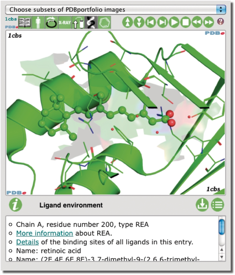
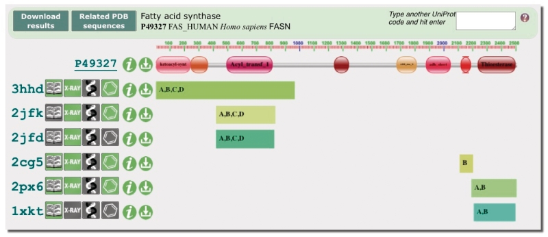
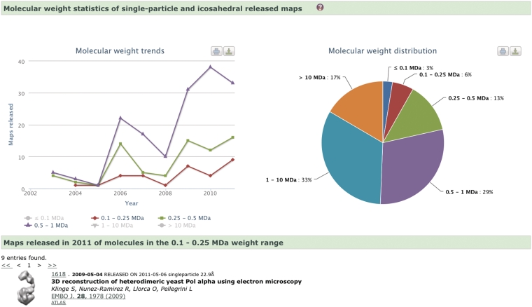
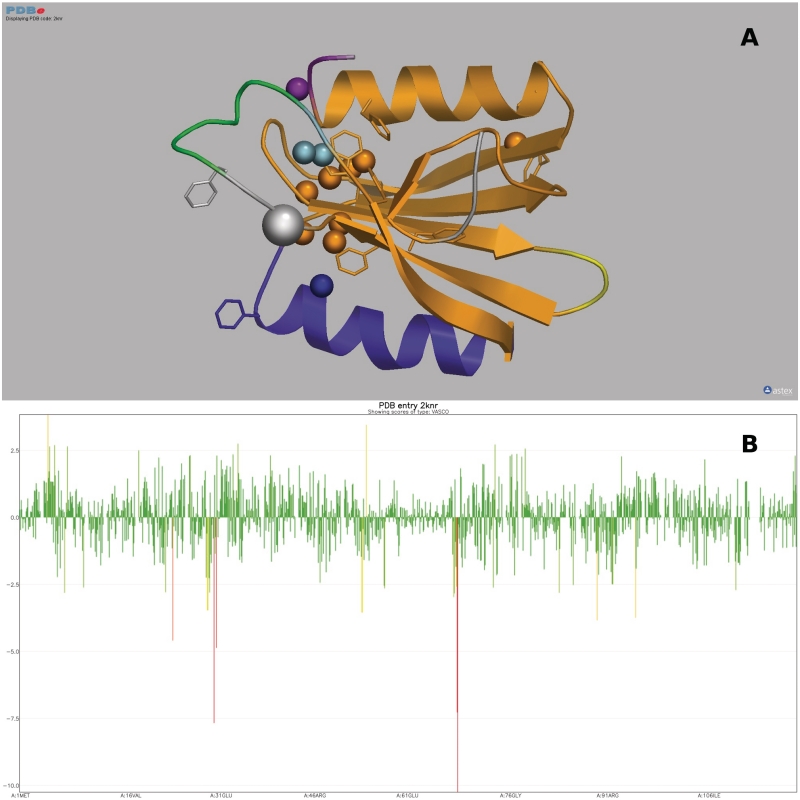
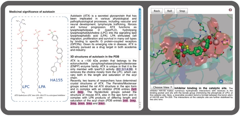
The Protein Data Bank in Europe (PDBe; pdbe.org) is a partner in the Worldwide PDB organization (wwPDB; wwpdb.org) and as such actively involved in managing the single global archive of biomacromolecular structure data, the PDB. In addition, PDBe develops tools, services and resources to make structure-related data more accessible to the biomedical community. Here we describe recently developed, extended or improved services, including an animated structure-presentation widget (PDBportfolio), a widget to graphically display the coverage of any UniProt sequence in the PDB (UniPDB), chemistry- and taxonomy-based PDB-archive browsers (PDBeXplore), and a tool for interactive visualization of NMR structures, corresponding experimental data as well as validation and analysis results (Vivaldi).


Figures

Figure 1.
Figure 1.
The PDBportfolio widget provides images and annotation about important aspects of one or more PDB entries, including the quaternary structure, domains and bound ligands. Refer to the text and pdbe.org/portfolio for more information, or see pdbe.org/1fcc for a live animated example.
Figure 2.
Figure 2.
The UniPDB widget provides a graphical overview of the sequence coverage of any UniProt entry in the PDB. This example (pdbe.org/unipdb?uniprot=FAS_HUMAN) shows the domain architecture of human fatty acid synthase (FAS), a multi-enzyme assembly line of fatty acids. Pfam identifies eight domains in human FAS, five of which have structural coverage in the PDB. Salient features of the relevant PDB entries are depicted using PDBlogos. Refer to the text and pdbe.org/unipdb for more information.
Figure 3.
Figure 3.
PDBeXpress (pdbe.org/express) can be used to find out which residues are observed to interact with a certain ligand in entries in the PDB. The graph shown here is for ATP and shows a preponderance of Gly, Lys, Ser and Thr residues, as expected in the Walker ATP-binding motif (21). The graph also shows other commonly observed residues in ATP binding sites such as Arg, Asp and Glu.
Figure 4.
Figure 4.
Molecular weight (MW) distribution and MW trends of the assemblies (single-particle and icosahedral) whose EM maps have been deposited in the EMDB archive, as generated with EMstats (pdbe.org/emstats). The distribution chart shows that only 22% of the studies are of assemblies with MW < 0.5 MDa. This is a consequence of the technical difficulties encountered when using the single-particle technique to study low-molecular-weight molecules or complexes. However, the trend charts show that the number of such studies is gradually increasing, which suggests that the field is learning how to overcome the experimental difficulties.
Figure 5.
Figure 5.
Use of Vivaldi to display and analyse PDB data about NMR entries [in this example, entry 2KNR which has been discussed by Lemak et al. (33): pdbe.org/vivaldi/2knr]. (A) Results of OLDERADO (22) and VASCO (23) analysis of the 2KNR ensemble presented in a 3D structure display. OLDERADO clusters the members of an ensemble of NMR models. The representative model of the largest cluster is shown as a cartoon. The colouring of the models signifies the various structural domains indentified by OLDERADO. This analysis indicates that while the central β-sheet and the N-terminal α-helix (all in orange) are rigid with respect to each other, the second α-helix (blue) forms a separate rigid body or ‘domain’. Atoms with unusual chemical shifts as identified by VASCO are shown as van der Waals spheres. In addition, aromatic residues are shown as sticks. This structure exhibits a number of chemical shift outliers that cluster together. However, many of these outliers are near aromatic rings, which are known to strongly affect the chemical shifts values of neighbouring atoms due to ring currents. (B) 1D graph of the deviation of chemical shift values from statistical averages as judged by VASCO as a function of residue number. The height and colour of the bars convey the extent of the deviation. This graph shows that most deviations are within usual bounds (green), but some of the outliers (yellow and red) are severe (up to 10 SD) and require a plausible explanation, such as the presence of a nearby aromatic ring.
Figure 6.
Figure 6.
Quips (pdbe.org/quips) is an interactive medium to develop stories about biomacromolecular structures. A Quips article contains links that activate the OpenAstexViewer applet to highlight and animate important facets of 3D structures. The text contains marked-up links to relevant literature, PDB entries, ligands, glossary terms, tutorials, further exploration options, etc. The example shown here (pdbe.org/quips?story=Autotaxin) accompanies a review on autotaxin structure and function (34). The review along with the interactive Quips provides users with better understanding of the topic when compared to static images.

References

    1. Bernstein FC, Koetzle TF, Williams GJ, Meyer EF, Jr, Brice MD, Rodgers JR, Kennard O, Shimanouchi T, Tasumi M. The Protein Data Bank: a computer-based archival file for macromolecular structures. J. Mol. Biol. 1977;112:535–542. - PubMed
    1. Berman HM. The Protein Data Bank: a historical perspective. Acta Crystallogr. 2008;A64:88–95. - PubMed
    1. Berman H, Henrick K, Nakamura H. Announcing the worldwide Protein Data Bank. Nat. Struct. Biol. 2003;10:980. - PubMed
    1. Berman H, Henrick K, Nakamura H, Markley JL. The worldwide Protein Data Bank (wwPDB): ensuring a single, uniform archive of PDB data. Nucleic Acids Res. 2007;35:D301–D303. - PMC - PubMed
    1. Rose PW, Beran B, Bi C, Bluhm WF, Dimitropoulos D, GoodsellD S, Prlic A, Quesada M, Quinn GB, Westbrook JD, et al. The RCSB Protein Data Bank: redesigned web site and web services. Nucleic Acids Res. 2011;39:D392–D401. - PMC - PubMed

Publication types