. 2012 Jan 15;28(2):198-205.
doi: 10.1093/bioinformatics/btr636. Epub 2011 Nov 21.

MetalionRNA: computational predictor of metal-binding sites in RNA structures

Affiliations

MetalionRNA: computational predictor of metal-binding sites in RNA structures

Anna Philips et al. Bioinformatics. .

Abstract

Motivation: Metal ions are essential for the folding of RNA molecules into stable tertiary structures and are often involved in the catalytic activity of ribozymes. However, the positions of metal ions in RNA 3D structures are difficult to determine experimentally. This motivated us to develop a computational predictor of metal ion sites for RNA structures.

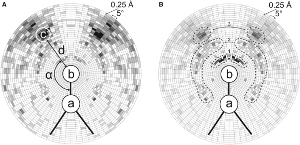
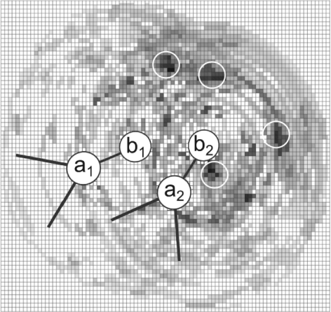
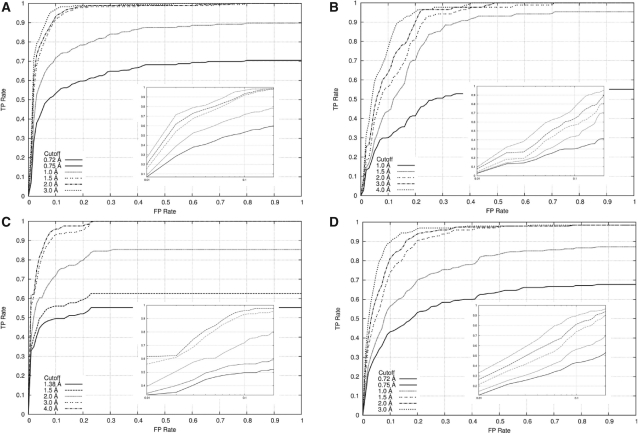
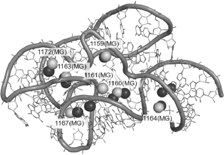
Results: We developed a statistical potential for predicting positions of metal ions (magnesium, sodium and potassium), based on the analysis of binding sites in experimentally solved RNA structures. The MetalionRNA program is available as a web server that predicts metal ions for RNA structures submitted by the user.

Availability: The MetalionRNA web server is accessible at http://metalionrna.genesilico.pl/.


Figures

Fig. 1.
Fig. 1.
Schematic views of the radial grid R used for deriving contact statistics for RNA atom pairs [a, b] in contact with cation c. The grid used for counting uses radial steps of 0.25Å and 5 around atom b (an O or N atom) and a (covalently bound to b). (A) Statistics of cation presence. For each cation, its distance d to the respective atom b, and the angle α (a, b, c) are calculated. The contact statistics derived for the RNA atom pair [P, OP2] and Mg2+ ions are shown in a gray scale (the more Mg2+ in a given bin, the darker the area). (B) The diagram shows the distribution of values for a normalized potential derived from contact statistics (A) for the RNA atom pair [P, OP2] and Mg2+ ions (the darker the area, the more negative value of the potential for the given bin). The three possible states of magnesium binding to RNA (Draper, 2004) are represented by the three peak tuples of the darkest areas. The first peak tuple (i) corresponds to Mg2+ chelated and partially dehydrated by phosphate groups of RNA. The second peak tuple (ii) corresponds to the water-mediated state. The third peak tuple (iii) represents the situation where the Mg2+ ion remains hydrated and interacts with the RNA via a layer of water molecules.
Fig. 2.
Fig. 2.
A schematic view of the cubic grid C used for deriving the potential for two [P, OP2] pairs. Only one layer of grid cells is represented and for simplicity we consider that all atoms are within this single layer. The potential W(n) is additive for cells <9Å from more than one OP2 atom. (the darker the area, the more negative value of the potential for the given grid cell). MetalionRNA places the center of a predicted cation in the grid cell with the lowest value, calculates the sum of W(n) of cells covered by the cation introduced and removes these cells from further consideration.
Fig. 3.
Fig. 3.
ROC curves to assess the classification performance of MetalionRNA with the width of 0.5Å for the cubic grid C using (A) the RNA-Mg2+ dataset, (B) the RNA-Na+ dataset, (C) the RNA−K+ dataset, (D) the DNA-Mg2+ dataset and various cut-off distance values (the maximum distance between a predicted and a real metal ion in which the prediction is marked as correct). In the big picture overall graph is shown, in the small picture only a range between 0 and 0.2Å is illustrated on a logarithmic scale.
Fig. 4.
Fig. 4.
Structure of the 23S rRNA fragment (PDB ID: 1HC8) with the experimentally determined positions of Mg2+ cations indicated by white labeled balls. Top-scoring Mg2+ cations predicted by MetalionRNA are shown as black balls. For detailed comparison of predicted and experimentally observed ions, see Table 2.

References

    1. Banatao D.R., et al. Microenvironment analysis and identification of magnesium binding sites in RNA. Nucleic Acids Res. 2003;31:4450–4460. - PMC - PubMed
    1. Berens C., et al. Visualizing metal-ion-binding sites in group I introns by iron(II)-mediated Fenton reactions. Chem. Biol. 1998;5:163–175. - PubMed
    1. Das R., Baker D. Automated de novo prediction of native-like RNA tertiary structures. Proc. Natl Acad. Sci. USA. 2007;104:14664–14669. - PMC - PubMed
    1. Draper D.E. A guide to ions and RNA structure. RNA. 2004;10:335–343. - PMC - PubMed
    1. Draper D.E. RNA folding: thermodynamic and molecular descriptions of the roles of ions. Biophys. J. 2008;95:5489–5495. - PMC - PubMed

Publication types