. 2010 Sep;19(2):75-82.
doi: 10.5607/en.2010.19.2.75. Epub 2010 Sep 30.

A winged-helix transcription factor foxg1 induces expression of mss4 gene in rat hippocampal progenitor cells

Affiliations

A winged-helix transcription factor foxg1 induces expression of mss4 gene in rat hippocampal progenitor cells

Ju-Suk Nam et al. Exp Neurobiol. 2010 Sep.

Abstract

Foxg1 (previously named BF1) is a winged-helix transcription factor with restricted expression pattern in the telencephalic neuroepithelium of the neural tube and in the anterior half of the developing optic vesicle. Previous studies have shown that the targeted disruption of the Foxg1 gene leads to hypoplasia of the cerebral hemispheres with severe defect in the structures of the ventral telencephalon. To further investigate the molecular mechanisms by which Foxg1 plays essential roles during brain development, we have adopted a strategy to isolate genes whose expression changes immediately after introduction of Foxg1 in cultured neural precursor cell line, HiB5. Here, we report that seventeen genes were isolated by ordered differential displays that are up-regulated by over-expression of Foxg1, in cultured neuronal precursor cells. By nucleotide sequence comparison to known genes in the GeneBank database, we find that nine of these clones represent novel genes whose DNA sequences have not been reported. The results suggest that these genes are closely related to developmental regulation of Foxg1.

Keywords: Foxg1; Mss4; ordered-differential display; telencephalon development.


Figures
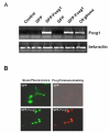
Fig. 1
Fig. 1
Detection of Foxg1 over-expressed in level of both mRNA and protein. The expression level of Foxg1 was increased in HiB5 cell line transfected with constructs encoding GFP or Foxg1 tagged with GFP by calcium-phosphate method. Twenty-four hours after transfection, cells were fixed or harvested and their total RNA was isolated. RT-PCR result indicated that mRNA level of Foxg1 was increased in HiB5 and R18F1 cells transfected with Foxg1 (A). mRNA from C6 cell line was used for positive control of Foxg1 and β-actin was used for internal control. Increased protein and cellular localization of Foxg1 were detected in HiB5 cell line transfected with Foxg1 by immunocytochemistry with anti-Foxg1 antibody (B). GFP was visualized as green fluorescence and Foxg1 was visualized as red fluorescence.
Fig. 2
Fig. 2
Example of the ordered differential display. Double-stranded cDNA was synthesized with non-extended ODD T-primer (5'-GCGAGTCGACCG(T)13) using double-stranded cDNA synthesis kit (Boehringer Mannheim) and the cDNA fragment pools, ligated with pseudo-double-stranded adaptor: long oligo (5'-GCGTGAAGACGACAGAAAGGGCGTGGTGCGGAGGGC GGT) and short one (5'-AC-CGCCCTCCGC), were amplified with adaptor-specific primer (5'-TGTAGCGTGAA GACGACAGAA) and non-extended ODD T-primer (A). Several primer combinations were used to amplify the cDNA fragments as described in materials and methods, and 33P-labeled PCR products were separated by PAGE (B). Each sample was prepared as duplication. Arrowheads indicate differential expressed bands.
Fig. 3
Fig. 3
Reverse-northern blot analysis of 17 confirmed cDNA fragments differentially expressed in Foxg1-transfected cells. Out of 100 Fragments, 17 were confirmed for their differential expression patterns. Note that the RNA for the cDNA probe was derived from another cell culture preparation than the one used for the original ODD. Arrows indicate the differentially expressed dots. Below the blots are the controls DNA, cyclophiline (CPN), demonstrating that equivalent amounts of total RNA were hybridized on each blots.
Fig. 4
Fig. 4
Expression of Mss4 in the heterozygote and homozygote Foxg1 mutant mouse brain. Sections from Foxg1(+/-) heterozygote (A) and Foxg1(-/-) mutant (B) at P0 were probed by in situ hybridization for Mss4 expression. cDNA fragment of Mss4 cloned in pCRII was used for dig-labeled probe by in vitro transcription. (A) A coronal section of the telencephalon from the Foxg1(+/-) heterozygote mice. (B) A coronal section of the telencephalon from the Foxg1(-/-) mutant mice. Thl: thalamus, Ctx: cortex.

Similar articles

Cited by

References

    1. Altschul SF, Madden TL, Schaffer AA, Zhang J, Zhang Z, Miller W, Lipman DJ. Gapped BLAST and PSI-BLAST: a new generation of protein database search programs. Nucleic Acids Res. 1997;25:3389–3402. - PMC - PubMed
    1. Ariani F, Hayek G, Rondinella D, Artuso R, Mencarelli MA, Spanhol-Rosseto A, Pollazzon M, Buoni S, Spiga O, Ricciardi S, Meloni I, Longo I, Mari F, Broccoli V, Zappella M, Renieri A. FOXG1 is responsible for the congenital variant of Rett syndrome. Am J Hum Genet. 2008;83:89–93. - PMC - PubMed
    1. Bourguignon C, Li J, Papalopulu N. XBF-1, a winged helix transcription factor with dual activity, has a role in positioning neurogenesis in Xenopus competent ectoderm. Development. 1998;125:4889–4900. - PubMed
    1. Burton JL, Burns ME, Gatti E, Augustine GJ, De Camilli P. Specific interactions of Mss4 with members of the Rab GTPase subfamily. EMBO J. 1994;13:5547–5558. - PMC - PubMed
    1. Danesin C, Peres JN, Johansson M, Snowden V, Cording A, Papalopulu N, Houart C. Integration of telencephalic Wnt and hedgehog signaling center activities by Foxg1. Dev Cell. 2009;16:576–587. - PubMed