. 2010 Sep;19(2):97-105.
doi: 10.5607/en.2010.19.2.97. Epub 2010 Sep 30.

Coordinate Regulation of Neurite Outgrowth by LRRK2 and Its Interactor, Rab5

Affiliations

Coordinate Regulation of Neurite Outgrowth by LRRK2 and Its Interactor, Rab5

Hye Young Heo et al. Exp Neurobiol. 2010 Sep.

Abstract

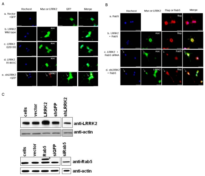
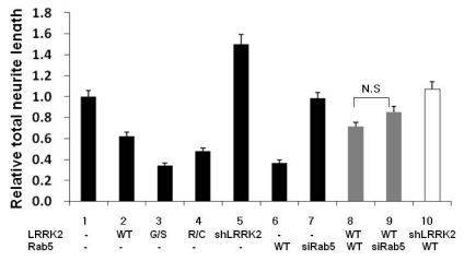
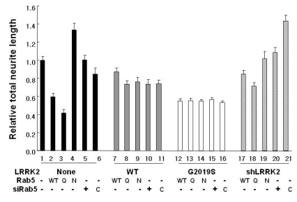
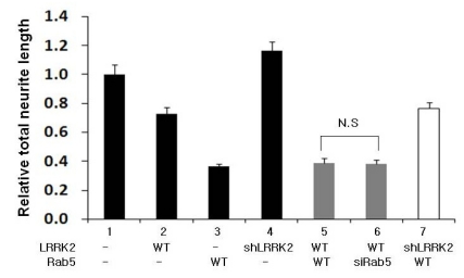
Neurite outgrowth and its maintenance are essential aspects of neuronal cells for their connectivity and communication with other neurons. Recent studies showed that over-expression of either leucine-rich repeat kinase 2 (LRRK2), whose mutations are associated with familial Parkinson's disease (PD), or Rab5b, an early endosomal marker protein, induces reduction in neurite length. Based on our recent findings that LRRK2 co-localizes and interacts with Rab5, we tested the hypothesis that LRRK2 and Rab5 may functionally interplay while controlling neurite outgrowth. Firstly, we confirmed previous reports that over-expression of either the LRRK2 PD-specific G2019S mutant or the Rab5 constitutively active Q79L mutant, but not of dominant negative N133I mutant, significantly reduces neurite outgrowth. Secondly, when over-expression of either LRRK2 wild type (WT) or G2019S was accompanied with over-expression of one of the Rab5 variants (WT, Q79L and N133I), or with down-regulation of Rab5, the reduction extent of its neurite length was similar to that of cells over-expressing LRRK2 alone, regardless of Rab5's status. Finally, we observed similar patterns of neurite length regulation in embryonic rat hippocampal neuron cultures. Taken together, our results suggest that LRRK2 and Rab5 functionally coordinate their regulation of neurite outgrowth and that LRRK2 is a more critical factor than Rab5.

Keywords: LRRK2; PC12 cells; Parkinson's disease; Rab5; neurite outgrowth.


Figures

Fig. 1
Fig. 1
(A) Representative fluorescence microscopic images of PC12 cells transfected with vector (a), myc tagged-LRRK2 wild type (b), -G2019S (c), -R1441C (d) and shLRRK2 (e) plasmids. (B) Representative fluorescence microscopic images of PC12 cells transfected with flag tagged Rab5 (a), myc tagged LRRK2 WT with flag tagged Rab5 plasmids (b), myc tagged LRRK2 WT with Rab5 siRNAs (c) and shLRRK2 with flag tagged Rab5 plasmids (d). PC12 cells were seeded on coverglass and treated with NGF (50 ng/ml) for 10 days. Cells were transfected with the indicated plasmid on the third day and fixed on the 10th day for microscopic analysis. Total amounts of DNA used were kept constant by addition of vector plasmids for all samples. For each case, nuclear staining was shown by Hoechst staining and proteins of interest were stained by the antibody against the tag (over-expression) or protein itself (down-expression). To identify cells down-expressing LRRK2 or Rab5, cells weakly stained by specific antibodies relative to adjacent cells were selected and used for analysis of neurite length (indicated by arrows). GFP plasmids were used to visualize neurites, if needed. The scale bar is 20µm. (C) Expression levels of LRRK2 and Rab5 in PC12 cells. PC12 cells were transfected by myc-LRRK2, flag-Rab5, shLRRK2 plasmids or Rab5 siRNA-1s (siRab5) and harvested for 3 or 2 days for LRRK2 or Rab5 analysis, respectively. The weak expression of LRRK2 and low transfection efficiency of the PC 12 cell line made it impossible to directly detect LRRK2 expression in western analysis. Therefore, the MACS (Miltenyi Biotec, Bergisch Gladbach, Germany) method was applied to sort positively transfected cells. PC12 cells transiently co-transfected with the indicated plasmids and pMACS4.1 expressing CD4Δ (Miltenyi Biotec) and sorted by application of magnetic field which detected cells bound to CD4 antibodies coupled to micromagnetic beads. Expression levels of both proteins are compared to the cellular actin level using anti-LRRK2, -Rab5 or -actin antibodies. *Indicates exogenously expressed flag tagged Rab5.
Fig. 2
Fig. 2
Analysis of the neurite lengths of PC12 cells transfected with the indicated plasmids and/or siRNAs. Cells were transfected and treated as explained in Fig. 1. Cells in five randomly selected fields were analyzed. Whether cells were transfected with the indicated plasmids and/or siRNAs was confirmed by the expression pattern of the proteins after staining with specific antibodies. Cells clearly showing over- or down-expression of corresponding proteins were selected. Every selected cell was subjected to neurite length analysis that was carried out using an automated image analysis program (MetaMorph Program, Molecular Devices, Downingtown, PA, USA) with the same parameter set for all samples. The resulting total neurite length of each cell is shown as average with standard error of mean (SEM). The Y axis is arbitrally set as 1 for the neurite length of cells transfected with vector plasmids. G/S and R/C indicate LRRK2 G2019S and R1441C mutants, respectively. N.S. means no significant difference between the indicated groups by ANOVA test (p>0.05). Lanes 2, 3, 4, 5, 6, 8 and 9, but not lanes 7 and 10, showed statistically significant differences compared to the control, lane 1 (p<0.05).
Fig. 3
Fig. 3
Neurite analysis of PC12 cells after over-expression of Rab5 wild type (WT), Q79L (Q) or N133I (N) with over- or down-expression of LRRK2. Total neurite length of each condition is shown as an average with SEM. All procedures were carried out as described in Fig. 2. C indicates GFP siRNAs used as a negative control.
Fig. 4
Fig. 4
Analysis of neurite length of rat hippocampal neuronal cells transfected with the indicated plasmids and/or siRNAs. Rat E17 primary dissociated hippocampal neuronal cells were cultured and the indicated plasmids and/or siRNAs were transiently transfected on the fourth day after seeding. Cells were fixed for microscopic analysis after incubation for 7 more days and analyzed for total neurite length as explained in Fig. 2. ns means no significant difference between the indicated groups by ANOVA test (p>0.05). Lanes 2, 3, 5, 6 and 7, but not lanes 4, showed statistically significant differences compared to the control, lane 1 (p<0.05).

References

    1. Dachsel JC, Taylor JP, Mok SS, Ross OA, Hinkle KM, Bailey RM, Hines JH, Szutu J, Madden B, Petrucelli L, Farrer MJ. Identification of potential protein interactors of Lrrk2. Parkinsonism Relat Disord. 2007;13:382–385. - PMC - PubMed
    1. Daitoku H, Isida J, Fujiwara K, Nakajima T, Fukamizu A. Dimerization of small GTPase Rab5. Int J Mol Med. 2001;8:397–404. - PubMed
    1. de Hoop MJ, Huber LA, Stenmark H, Williamson E, Zerial M, Parton RG, Dotti CG. The involvement of the small GTP-binding protein Rab5a in neuronal endocytosis. Neuron. 1994;13:11–22. - PubMed
    1. Deng J, Lewis PA, Greggio E, Sluch E, Beilina A, Cookson MR. Structure of the ROC domain from the Parkinson's disease-associated leucine-rich repeat kinase 2 reveals a dimeric GTPase. Proc Natl Acad Sci U S A. 2008;105:1499–1504. - PMC - PubMed
    1. Gandhi PN, Chen SG, Wilson-Delfosse AL. Leucine-rich repeat kinase 2 (LRRK2): a key player in the pathogenesis of Parkinson's disease. J Neurosci Res. 2009;87:1283–1295. - PMC - PubMed