. 2011 Mar;20(1):45-53.
doi: 10.5607/en.2011.20.1.45. Epub 2011 Mar 31.

Rapid Disruption of Cellular Integrity of Zinc-treated Astroglia Is Regulated by p38 MAPK and Ca-dependent Mechanisms

Affiliations

Rapid Disruption of Cellular Integrity of Zinc-treated Astroglia Is Regulated by p38 MAPK and Ca-dependent Mechanisms

Joo-Young Im et al. Exp Neurobiol. 2011 Mar.

Abstract

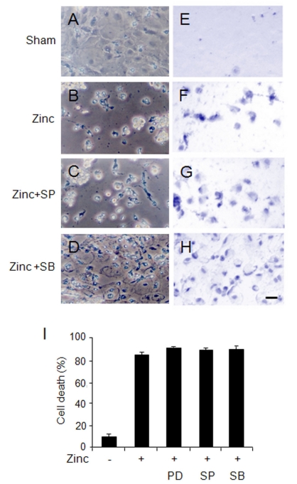
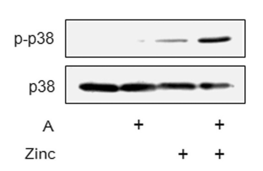
Cultured cortical primary astroglia treated with zinc died while rapidly detached from culture plates, a distinct part of zinc-treated astroglia. In the present study, we investigated the mechanism underlying the rapid change in the morphologic integrity of zinc-treated astroglia. Among the early cellular events occurring in zinc-treated astroglia, strong activation of p38 MAPK and JNK was evident. Although inhibitors of p38 (SB203580 and SB202190) or JNK (SP600125) did not protect zinc-insulted astroglia from cell death, the p38 inhibitors, but not the JNK inhibitor, suppressed actin filament and cell morphology disruption. The Ca(2+) ionophore, A23187, also suppressed actin filament and cell morphology disruption, but not cell death, of zinc-insulted astroglia. However, A23187 did not inhibit p38 MAPK activation in zinc-treated astroglia. Together these results suggest that zinc influx in astroglia results in rapid loss of the morphologic integrity via mechanisms regulated by p38 kinase and/or Ca(2+) signaling.

Keywords: actin filament; astroglia; morphology protection; p38 inhibitors; zinc.


Figures

Fig. 1
Fig. 1
p38 and JNK MAPKs were activated in zinc-treated astroglia. (A, B) Astroglial cell death by zinc increased in a zinc-dose (A) and zinc-treated time (B) dependent manner. LDH assay was performed after 24 h-exposure of cultures to indicated concentrations of zinc. (C) Western blots showing zinc-dose dependent activation of p38 and JNK MAPKS in astroglia. Assay was performed 3 h after zinc treatment. Anti-phospho-p38 MAPK and anti-phospho-JNK were used to deduce the activation levels of these kinases. (D) Western blots showing time-dependent activation of p38 and JNK MAPKS in zinc (35 µM)-treated astroglia.
Fig. 2
Fig. 2
The p38 MAPK inhibitor, SB203580, suppressed zinc-induced morphology disruption and detachment from the culture plate. (A~H) Phase contrast (A~D) or trypan-blue stained (E~H) astroglia with sham treatment (A, E) or astroglia treated with zinc (B, F), with zinc and SP600125 (C, G), or with zinc and SB203580 (D, H). Photomicrographs were taken 24 h after zinc (35 µM)-treatment in the presence of SP600125 (20 µM) or SB203580 (20 µM). Scale bar represents 100 µm. (I) LDH assay showing cell death levels of zinc (35 µM)-treated astroglia in the presence of PD98059 (PD: 20 µM), SP600125 (SP: 20 µM), or SB203580 (SB: 20 µM). LDH assay was performed 24 h after zinc treatment.
Fig. 3
Fig. 3
The Ca2+ ionophore, A23187, suppressed cell morphology disruption of zinc-treated astroglia. (A) LDH assay showing cell death levels of astroglia treated with A23187 (0.1 µ M), zinc (35 µM), zinc (35 µM)+A23187 (0.1 µM), or zinc (35 µM)+SB203580 (20 µM). LDH assay was performed after 24 h of drug treatment. (A) A23187; SB, SB203580. (B~E) Trypan-blue stained astroglia treated with zinc (35 µM), zinc (35 µM)+SB203580 (20 µM), A23187 (0.1 µM) or zinc (35 µM)+A23187. Photomicrographs were taken after 24 h of zinc treatment in the presence of SB203580 or A23187. (F~K) Trypan-blue stained astroglia with sham treatment (F, G), with zinc (H, I), or with zinc and SB203580 (J, K) in the presence or absence of the Ca2+ chelator, BAPTA (10 µM). Photomicrographs were taken 24 h after treatment of zinc (35 µM) and SB203580 (20 µM). (L) LDH assay showing cell death levels of astroglia treated with zinc (35 µM) or zinc (35 µM)+SB203580 (20 µM) in the presence or absence of BAPTA (10 µM). LDH assay was performed after 24 h of drug treatment. Scale bar represents 100 µm. SB, SB203580. The data are presented as means±S.E.M. (n=6~12). **Denotes difference at p<0.05.
Fig. 4
Fig. 4
SB203580 and A23187 suppressed actin-filament disruption in zinc-treated astroglia. (A~E) Photomicrographs showing actin-filament distributions in zinc-treated astroglia stained with TRITC-labeled phalloidin. Sham control (A) and astroglia treated with Zn (35 µM) alone (B), Zn (35 µM)+SB203580 (20 µM) (C), A23187 (0.1 µM) alone (D), or zinc (35 µM)+A23187 (0.1 µM) (E). Actin-filament disruptions were visualized after 10 h of zinc treatment. Scale bar represents 100 µm.
Fig. 5
Fig. 5
A23187 did not suppress p38 MAPK activation in zinc-treated astroglia. Western blots showing p38 MAPK activation levels in zinc-treated astroglia. Western blot analysis was performed after 3 h of treatment with zinc (40 µM) alone or zinc (40 µM)+A23187 (A: 0.1 µM).

References

    1. An WL, Bjorkdahl C, Liu R, Cowburn RF, Winblad B, Pei JJ. Mechanism of zinc-induced phosphorylation of p70 S6 kinase and glycogen synthase kinase 3beta in SH-SY5Y neuroblastoma cells. J Neurochem. 2005;92:1104–1115. - PubMed
    1. Arteaga S, Andrade-Cetto A, Cardenas R. Larrea tridentata (creosote bush), an abundant plant of Mexican and U.S. American deserts and its metabolite nordihydroguaiaretic acid. J Ethnopharmacol. 2005;98:231–239. - PubMed
    1. Carlier MF, Valentin-Ranc C, Combeau C, Fievez S, Pantoloni D. Actin polymerization: regulation by divalent metal ion and nucleotide binding, ATP hydrolysis and binding of myosin. Adv Exp Med Biol. 1994;358:71–81. - PubMed
    1. Carragher NO, Frame MC. Calpain: a role in cell transformation and migration. Int J Biochem Cell Biol. 2002;34:1539–1543. - PubMed
    1. Chae HJ, Ha HY, Im JY, Song JY, Park S, Han PL. JSAP1 is required for the cell adhesion and spreading of mouse embryonic fibroblasts. Biochem Biophys Res Commun. 2006;345:809–816. - PubMed