. 2011 Nov;9(11):e1001196.
doi: 10.1371/journal.pbio.1001196. Epub 2011 Nov 15.

Cryo-electron tomography of Marburg virus particles and their morphogenesis within infected cells

Affiliations

Cryo-electron tomography of Marburg virus particles and their morphogenesis within infected cells

Tanmay A M Bharat et al. PLoS Biol. 2011 Nov.

Abstract

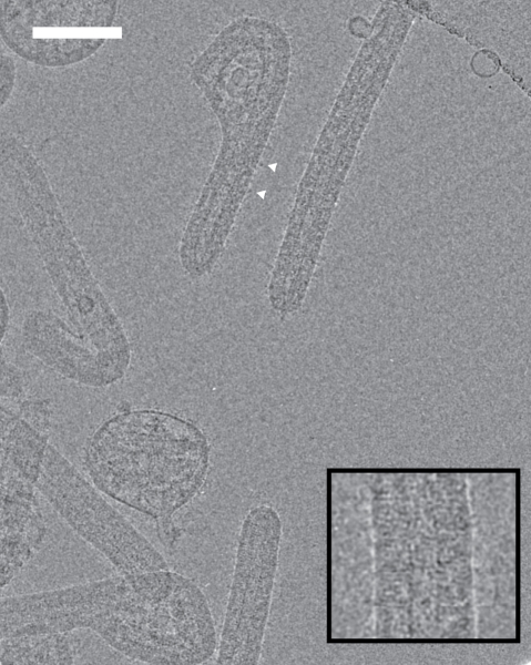
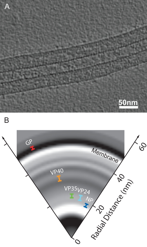
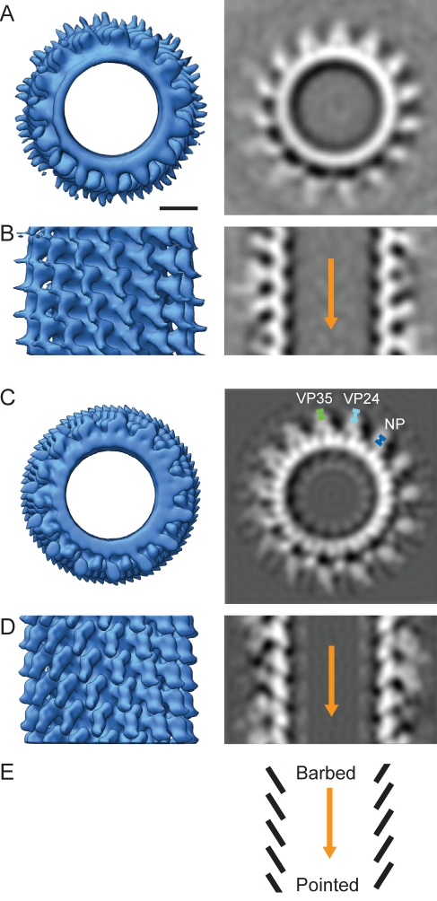
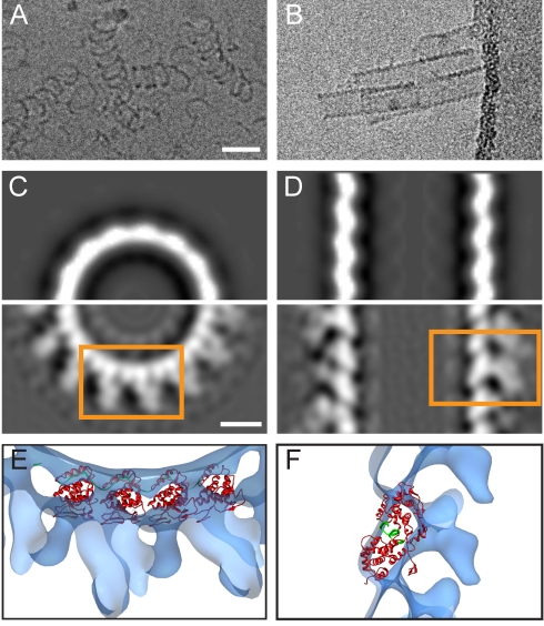
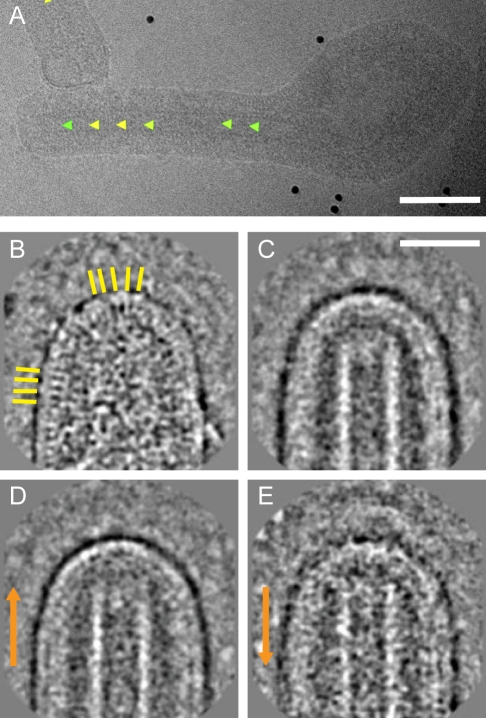
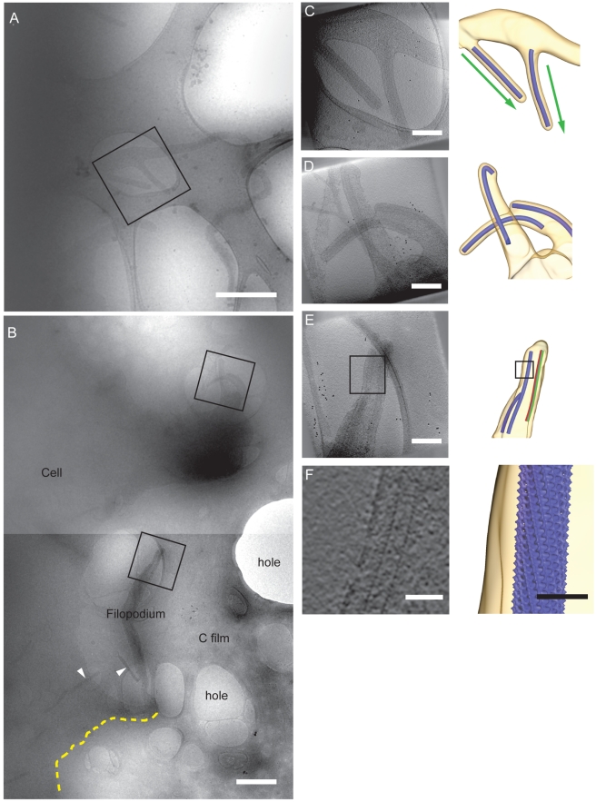
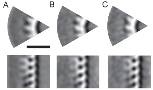
Several major human pathogens, including the filoviruses, paramyxoviruses, and rhabdoviruses, package their single-stranded RNA genomes within helical nucleocapsids, which bud through the plasma membrane of the infected cell to release enveloped virions. The virions are often heterogeneous in shape, which makes it difficult to study their structure and assembly mechanisms. We have applied cryo-electron tomography and sub-tomogram averaging methods to derive structures of Marburg virus, a highly pathogenic filovirus, both after release and during assembly within infected cells. The data demonstrate the potential of cryo-electron tomography methods to derive detailed structural information for intermediate steps in biological pathways within intact cells. We describe the location and arrangement of the viral proteins within the virion. We show that the N-terminal domain of the nucleoprotein contains the minimal assembly determinants for a helical nucleocapsid with variable number of proteins per turn. Lobes protruding from alternate interfaces between each nucleoprotein are formed by the C-terminal domain of the nucleoprotein, together with viral proteins VP24 and VP35. Each nucleoprotein packages six RNA bases. The nucleocapsid interacts in an unusual, flexible "Velcro-like" manner with the viral matrix protein VP40. Determination of the structures of assembly intermediates showed that the nucleocapsid has a defined orientation during transport and budding. Together the data show striking architectural homology between the nucleocapsid helix of rhabdoviruses and filoviruses, but unexpected, fundamental differences in the mechanisms by which the nucleocapsids are then assembled together with matrix proteins and initiate membrane envelopment to release infectious virions, suggesting that the viruses have evolved different solutions to these conserved assembly steps.


Conflict of interest statement

The authors have declared that no competing interests exist.

Figures

Figure 1
Figure 1. CryoEM of MARV.
CryoEM image of the morphologies observed in MARV samples. Scale bar, 100 nm, electron density black. Arrowheads indicate examples of spike-like membrane protrusions. Inset: magnified view of part of a filamentous particle showing striations under the membrane.
Figure 2
Figure 2. CryoET of MARV and mapping of viral proteins.
(A) A slice through a tomogram showing a section of a MARV virion. Electron density is black. (B) The radial positions of the proteins in the virion, represented by the mean, Gaussian-corrected positions of the gold beads in IEM have been superimposed onto a radial density slice of the virions produced from the aligned subtomograms (Text S1). The length of the bar represents the error associated with each IEM measurement. Electron density is white. See Figure S1 and Table S1 for further details.
Figure 3
Figure 3. Reconstruction of the MARV NC.
(A–B) Reconstruction of the MARV NC from cryoET and subtomogram averaging. (A) The NC helix is shown viewed along the helical axis as an isosurface (left) and as a section through the density (right). All isosurfaces are displayed at a contour level of 1.5 σ away from the mean. Scale bar, 10 nm. Electron density is white. (B) Side-on view of helix. The orange arrow is directed towards the pointed end of the NC helix. (C–D) Reconstruction of the MARV NC from 2-D helical reconstruction techniques in the same orientations as (A–B). The radial positions of the NC proteins as determined from the mean location of the gold beads earlier in Figure 2B have been superimposed (C, right). (E) Schematic representation of the NC highlighting the “pointed” and “barbed” end of the helix. See Figures S2 and S3.
Figure 4
Figure 4. Assignment of the core region of MARV NP and fitting of VSV NP into the MARV NC density.
(A) CryoEM image of MARV NP purified from 293T cells. Scale bar, 50 nm, electron density black. (B) CryoEM image of MARV NP(1–390) produced in a parallel experiment. (C–D) Comparison of the MARV NC reconstruction in Figure 2 with that of the MARV NP(1–390) helix. (C) A section perpendicular to the helical axis for the NC helix (lower panel) compared to the corresponding part from the NPΔ390 helix (upper panel). Scale bar, 10 nm, electron density white. (D) Corresponding section along the helical axis. (E–F) Fitting of the pseudo-atomic model of the VSV NP (PDB 2WYY with protein in red and RNA in green) into the full MARV NC reconstruction, after assignment of the NP density. The views shown correspond to the regions within the orange boxes in (C–D).
Figure 5
Figure 5. Directionality of the NC in free virions and structure of MARV tips.
(A) A six-shaped MARV with arrowheads indicating the “pointed end” orientation assigned during helical reconstruction. Color represents cross-correlation coefficient (green∶high, yellow∶low) between extracted boxes and corresponding reprojections of the NC reconstruction. Scale bar, 100 nm, electron density black. (B) A representative extracted “pointed” MARV tip. Yellow lines highlight VP40 striations. Scale bar, 50 nm, electron density white. (C) Two-dimensional average of all extracted MARV tips. (D) Average of all “pointed” MARV tips. (E) Average of all “barbed” MARV tips. Orange arrows are directed towards the pointed end of the NC.
Figure 6
Figure 6. CryoET of MARV budding from infected cells.
(A,B) Low magnification images of MARV budding from infected cells. Areas selected as targets for tomography are marked with a black box. The carbon film and examples of holes in the film, the cell, and a filopodium are annotated. NCs in free or budding viruses are marked with arrowheads. Scale bar, 1 µm, electron density black. (C–E) Tomographic reconstructions of targeted regions. Left: projection taken through the reconstructed tomogram. Right: isosurface rendering of the same region. Membrane is shown in yellow and the NC as a blue cylinder. The NC direction, as determined by subtomogram averaging, is indicated by the arrows. An example of membrane-associated (green) and free (red) sides of the NC used for subtomogram averaging is shown in (E). Scale bars, 200 nm. Animations of these tomograms are included as Movies S2, S3, S4. (F) Higher magnification of regions demarcated by boxes in (E) as a slice through the tomogram (left), showing repeating features corresponding to the inner NC helix and as an isosurface representation (right), created by placing the NC reconstruction back into the tomogram. Scale bars, 50 nm.
Figure 7
Figure 7. 3-D reconstruction of the budding MARV NC.
(A) Slice perpendicular to (top) and along (bottom) the NC axis showing a segment of the subtomogram averaging reconstruction of fully enveloped budding MARV NCs (class I). (B) Reconstruction of the membrane associated side of budding NCs that were not fully enveloped on all sides (class II). (C) Corresponding reconstruction of the non-membrane-associated NC (class III).

Comment in

Similar articles

Cited by

References

    1. Lamb R. Mononegavirales. In: Knipe D, Howley P, editors. Fields Virology. Fifth Edition ed. Philadelphia, USA: Lippincott Williams and Wilkins; 2007. 1357
    1. Albertini A, Schoehn G, Weissenhorn W, Ruigrok R. Structural aspects of rabies virus replication. Cell Mol Life Sci. 2008;65:282–294. - PMC - PubMed
    1. Luo M, Green T, Zhang X, Tsao J, Qiu S. Structural comparisons of the nucleoprotein from three negative strand RNA virus families. Virol J. 2007;4:72. - PMC - PubMed
    1. Desfosses A, Goret G, Estrozi L. F, Ruigrok R. W, Gutsche I. Nucleoprotein-RNA orientation in the measles virus nucleocapsid by three-dimensional electron microscopy. J Virol 2010 - PMC - PubMed
    1. Ge P, Tsao J, Schein S, Green T. J, Luo M, et al. Cryo-EM model of the bullet-shaped vesicular stomatitis virus. Science. 2010;327:689–693. - PMC - PubMed

Publication types

MeSH terms