

Cholangiocarcinoma in magnetic resonance cholangiopancreatography and fascioliasis in endoscopic ultrasonography
- PMID: 22110417
- PMCID: PMC3219480
- DOI: 10.1159/000333229
Cholangiocarcinoma in magnetic resonance cholangiopancreatography and fascioliasis in endoscopic ultrasonography
Abstract
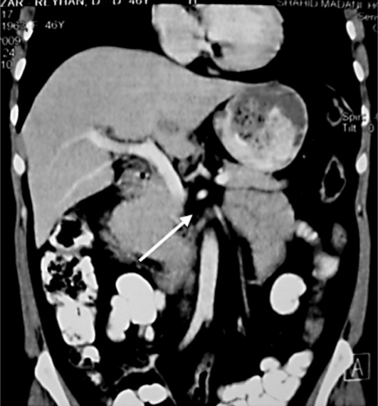
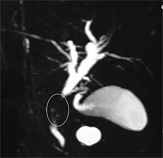
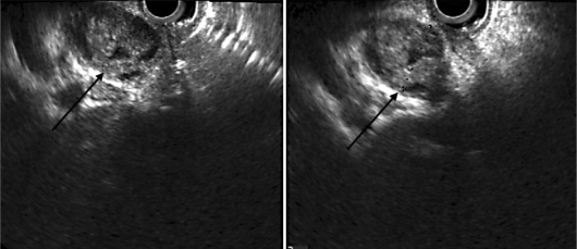
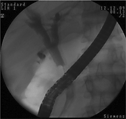
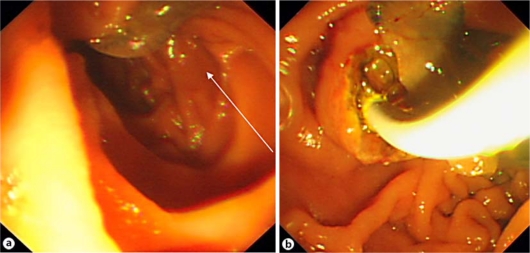
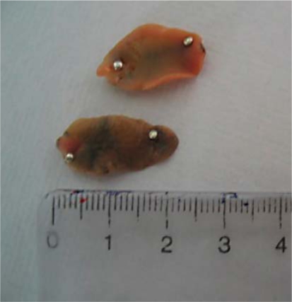
Fascioliasis is a worldwide zoonotic infection with Fasciola hepatica and Fasciola gigantica. The zoonoses are particularly endemic in sheep-raising countries and are also endemic in Iran. Typical symptoms that may be associated with fascioliasis can be divided by phases of the disease, including the acute or liver phase, the chronic or biliary phase, and ectopic or pharyngeal fascioliasis. Cholestatic symptoms may be absent, and in some cases diagnosis and treatment may be preceded by a long period of abdominal pain, eosinophilia and vague gastrointestinal symptoms. We report a case with epigastric and upper quadrant abdominal pain for the last 4 years, with imaging suggesting cholangiocarcinoma. Considering a new concept of endoscopic ultrasonography, at last F. hepatica was extracted with endoscopic retrograde cholangiography.
Keywords: Endoscopic retrograde cholangiopancreatography; Endoscopic ultrasonography; Fasciola hepatica; Magnetic resonance cholangiopancreatography.
Figures
Similar articles
-
Finding of biliary fascioliasis by endoscopic ultrasonography in a patient with eosinophilic liver abscess.Case Rep Gastroenterol. 2014 Oct 8;8(2):310-8. doi: 10.1159/000367592. eCollection 2014 Sep. Case Rep Gastroenterol. 2014. PMID: 25473389 Free PMC article.
-
Endoscopic management of biliary fascioliasis: a case report.J Med Case Rep. 2010 Mar 6;4:83. doi: 10.1186/1752-1947-4-83. J Med Case Rep. 2010. PMID: 20205932 Free PMC article.
-
A case of Fasciola hepatica infection mimicking cholangiocarcinoma and ITS-1 sequencing of the worm.Korean J Parasitol. 2014 Apr;52(2):193-6. doi: 10.3347/kjp.2014.52.2.193. Epub 2014 Apr 18. Korean J Parasitol. 2014. PMID: 24850964 Free PMC article.
-
Fascioliasis: a worldwide parasitic disease of importance in travel medicine.Travel Med Infect Dis. 2014 Nov-Dec;12(6 Pt A):636-49. doi: 10.1016/j.tmaid.2014.09.006. Epub 2014 Sep 28. Travel Med Infect Dis. 2014. PMID: 25287722 Review.
-
[Endoscopic removal by ERCP of Fasciola hepatica alive: two case reports and review of the literature].Rev Gastroenterol Peru. 2013 Jan-Mar;33(1):75-81. Rev Gastroenterol Peru. 2013. PMID: 23539060 Review. Spanish.
Cited by
-
Finding of biliary fascioliasis by endoscopic ultrasonography in a patient with eosinophilic liver abscess.Case Rep Gastroenterol. 2014 Oct 8;8(2):310-8. doi: 10.1159/000367592. eCollection 2014 Sep. Case Rep Gastroenterol. 2014. PMID: 25473389 Free PMC article.
-
New features of fascioliasis in human and animal infections in Ilam province, Western Iran.Gastroenterol Hepatol Bed Bench. 2013 Summer;6(3):152-5. Gastroenterol Hepatol Bed Bench. 2013. PMID: 24834263 Free PMC article.
-
Biliary fascioliasis--an uncommon cause of recurrent biliary colics: report of a case and brief review.Ger Med Sci. 2012;10:Doc10. doi: 10.3205/000161. Epub 2012 May 2. Ger Med Sci. 2012. PMID: 22566787 Free PMC article. Review.
-
Oxysterols of helminth parasites and pathogenesis of foodborne hepatic trematodiasis caused by Opisthorchis and Fasciola species.Parasitol Res. 2020 May;119(5):1443-1453. doi: 10.1007/s00436-020-06640-4. Epub 2020 Mar 23. Parasitol Res. 2020. PMID: 32206886 Review.
-
Fasciola Infection Unexpectedly Found During Cholecystectomy: Review on How to Avoid Increasing Surgery Interventions in Non-human Endemic Areas.Acta Parasitol. 2023 Dec;68(4):891-902. doi: 10.1007/s11686-023-00726-6. Epub 2023 Nov 7. Acta Parasitol. 2023. PMID: 37934346 Free PMC article.
References
-
- Gulsen M, Savas MC, Koruk M, Kadayifci A, Demirci F. Fascioliasis: a report of five cases presenting with common bile duct obstruction. Neth J Med. 2006;64:17–19. - PubMed
-
- Aksoy DY, Kerimoglu U, Oto A, et al. Infection with Fasciola hepatica. Clin Microbiol Infect. 2005;11:859–861. - PubMed
-
- Condomines J, Rene-Espinet JM, Espinos-Perez JC, Vilardell F. Percutaneous cholangiography in the diagnosis of hepatic fascioliasis. Am J Gastroenterol. 1985;80:384–386. - PubMed
-
- Haseeb AN, el-Shazly AM, Arafa MA, Morsy AT. A review on fascioliasis in Egypt. J Egypt Soc Parasitol. 2002;32:317–354. - PubMed
-
- S Mas-Coma. Epidemiology of fascioliasis in human endemic areas. J Helminthol. 2005;79:207–216. - PubMed
Publication types
LinkOut - more resources
Full Text Sources
