Case Reports
. 2011;6(11):e26159.
doi: 10.1371/journal.pone.0026159. Epub 2011 Nov 16.

SNCA triplication Parkinson's patient's iPSC-derived DA neurons accumulate α-synuclein and are susceptible to oxidative stress

Affiliations
Case Reports

SNCA triplication Parkinson's patient's iPSC-derived DA neurons accumulate α-synuclein and are susceptible to oxidative stress

Blake Byers et al. PLoS One. 2011.

Abstract

Parkinson's disease (PD) is an incurable age-related neurodegenerative disorder affecting both the central and peripheral nervous systems. Although common, the etiology of PD remains poorly understood. Genetic studies infer that the disease results from a complex interaction between genetics and environment and there is growing evidence that PD may represent a constellation of diseases with overlapping yet distinct underlying mechanisms. Novel clinical approaches will require a better understanding of the mechanisms at work within an individual as well as methods to identify the specific array of mechanisms that have contributed to the disease. Induced pluripotent stem cell (iPSC) strategies provide an opportunity to directly study the affected neuronal subtypes in a given patient. Here we report the generation of iPSC-derived midbrain dopaminergic neurons from a patient with a triplication in the α-synuclein gene (SNCA). We observed that the iPSCs readily differentiated into functional neurons. Importantly, the PD-affected line exhibited disease-related phenotypes in culture: accumulation of α-synuclein, inherent overexpression of markers of oxidative stress, and sensitivity to peroxide induced oxidative stress. These findings show that the dominantly-acting PD mutation is intrinsically capable of perturbing normal cell function in culture and confirm that these features reflect, at least in part, a cell autonomous disease process that is independent of exposure to the entire complexity of the diseased brain.


Conflict of interest statement

Competing Interests: The authors have declared that no competing interests exist.

Figures

Figure 1
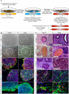
Figure 1. Analysis of pluripotency of iPSC lines.
(A) Human dermal fibroblasts (HDF) from Trpl17 and Ctrl2 were reprogrammed into iPSCs capable of forming colonies on mouse embryonic fibroblasts (MEFs) that expressed pluripotency associated antigens, SSEA3, TRA1-60, TRA-181, NANOG, and SSEA4 and were negative for lineage marker SSEA1. (B) Pluripotency of iPSC lines Trpl17 and Ctrl2 was assayed through immunohistochemistry of in vivo teratoma formation, demonstrating formation of cell types from all three germ layers; epithelial tissue (endoderm), cartilage (mesoderm) and neuronal tissue (ectoderm). Pluripotency potential was further characterized through embryoid body differentiation in vitro. Immunofluorescent staining confirmed the presence of cell types indicative of all three germ layers, βIII-tubulin and TH (ectoderm), desmin and α-SMA (mesoderm) and a-fetoprotein (endoderm; Figure S4A).
Figure 2
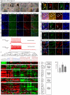
Figure 2. Characterization of directed dopaminergic neuron differentiation.
(A) Trpl17 and Ctrl2 neural induction time course 5× phase contrast images at iso-location (except where noted): Days 3, 6, 9 in the upper row are images of the same location in each culture. However, rosettes formed on the expanding colony periphery and not at the initial site of colony adhesion. Thus, the lower row shows days 9, 12, and 14 at a different location in each culture. Formations of neural rosettes (example in white circle) were visible begging on day 9 and continued to expand and proliferate through day 14 and beyond. (B) IF analysis of pluripotent lines H9, Trpl17, and Ctrl2 for DAPI, Nestin, DCX and TH. The presence of significant numbers of TH positive neurons demonstrates the ability to differentiate and enrich for dopaminergic neurons. IF confocal micrograph demonstrating (C) midbrain specific transcription factor pitx3 colocalization with TH and SNCA in Trpl17 dopaminergic neurons; (D) midbrain associated ion channel GIRK2, (E) midbrain specific transcription factor EN1, and (F) midbrain associated transcription factor LMX1a were expressed in Trpl17 and Ctrl2 dopaminergic neurons. (G) FOXA2, a marker expressed by floor plate progenitors and their midbrain dopaminergic neuron derivatives, was expressed in Trpl17 and Ctrl2 dopaminergic neurons. (H) Single cell patch-clamp recordings demonstrate that neurons derived from Trpl17 iPSCs are electrophysiologically functional with multiple spike trains and a normal −70 mV resting potential. (I) Clustering of samples and genes in a 96×96 microfluidic qPCR array. Samples clustered by differentiation stage (undifferentiated, rosette and DA neurons) rather than by line (H9, Trpl8, Trpl17, Trpl43, Ctrl1, Ctrl2, or Ctrl3.). Fetal brain samples clustered with DA neurons rather than undifferentiated or rosettes cells. Five self-clustered gene sections, of the full 96 genes (Figure S7), were examined closely, demonstrating self-assembly of pluripotency, general neural, and mature neural related genes with expression patterns that correlate to sample developmental stage and human fetal brain samples. (J) The number of TH positive cells per well was not significantly different, although Trpl17 had a greater variance. Scale bars = 100 µm (a–b), 10 µm (c, e), 25 µm (d, f–g) and error bars = S.E.M.
Figure 3
Figure 3. Characterization of midbrain DA neurons and phenotypic differences between pluripotent lines.
(A) IF confocal micrographs of DAPI, α-synuclein, TH, and DCX, demonstrate variable α-synuclein accumulation in Trpl17 derived DA neurons. (B) SNCA gene copy number computation through qPCR of SNCA 3rd intron. Absolute copy number (2×2−ΔCT) is reported: Ctrl-HDF, Ctrl2, and H9 ES cells all had a normal diploid copy number. Trpl-HDF and Trpl17 possessed 4 allelic copies of SNCA, consistent with a single allele triplication. (C) SNCA mRNA expression analysis shows an ANOVA significant difference between all three lines, and a post-hoc significant SNCA expression difference between Trpl17 and H9 neurons. (D) Western blots show greater abundance of 17 kDa monomeric α-synuclein in Trpl17 DA cultures than control Ctrl2 and H9. Cells from two independently differentiated wells are shown per sample. (E) The ratio of monomeric α-synuclein/GAPDH was calculated for each sample and results are expressed as the mean of two samples. Scale bar = 50 µm. Error bars = SEM in c and d and sample range in g. *p<0.05 and **p<0.001.
Figure 4
Figure 4. Analysis of oxidative stress levels and response to peroxide induced cell stress.
(A–E) ANOVA significance analysis across Ctrl2, Trpl17 and H9 cultures revealed five differentially expressed stress related genes. Trpl17 neural cultures had the highest expression of each ANOVA identified differentially expressed stress related gene; DNAJA1, HMOX2, UCHL1, HSPB1 and MAO-A. (F) Confocal micrographs showing activated Caspase-3 in Trpl17 DA cultures with and without H2O2 treatment. (G) A plot of the fraction of TH+ cells that were activated Caspase-3 positive for each line with and without 200 µM H2O2 treatment. Error bars = S.E.M. and scale bars = 100 µm (j) and 10 µm (k). *p<0.05, **p<0.001.

References

    1. Hughes AJ, Daniel SE, Kilford L, Lees AJ. Accuracy of clinical diagnosis of idiopathic Parkinson's disease: a clinico-pathological study of 100 cases. J Neurol Neurosurg Psychiatry. 1992;55:181–184. - PMC - PubMed
    1. Wakabayashi K, Takahashi H. Neuropathology of autonomic nervous system in Parkinson's disease. Eur Neurol. 1997;38(Suppl 2):2–7. - PubMed
    1. Langston JW. The Parkinson's complex: parkinsonism is just the tip of the iceberg. Ann Neurol. 2006;59:591–596. - PubMed
    1. Lewy F. Paralysis agitans. In: Lewandowsky M, editor. Pathologische Anatomie Handbuch der Neurologie. Berlin: Springer-Verlag; 1912. 13
    1. Lewy F. Zur pathologischen Anatomie der Paralysis Agitans. Deutsche Zeitschrift fur Nervenheilkunde. 1913;50:50–55.

Publication types

MeSH terms