. 2011;6(11):e26803.
doi: 10.1371/journal.pone.0026803. Epub 2011 Nov 16.

Long circulating lectin conjugated paclitaxel loaded magnetic nanoparticles: a new theranostic avenue for leukemia therapy

Affiliations

Long circulating lectin conjugated paclitaxel loaded magnetic nanoparticles: a new theranostic avenue for leukemia therapy

Abhalaxmi Singh et al. PLoS One. 2011.

Abstract

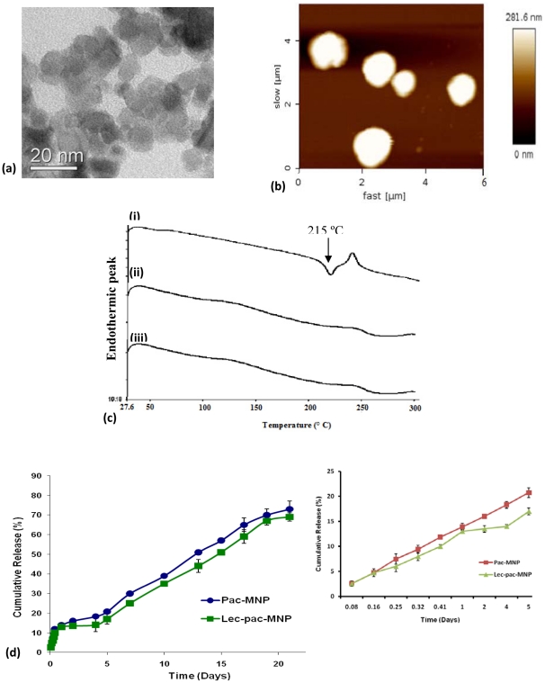
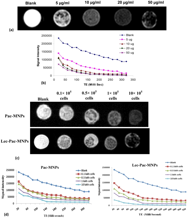
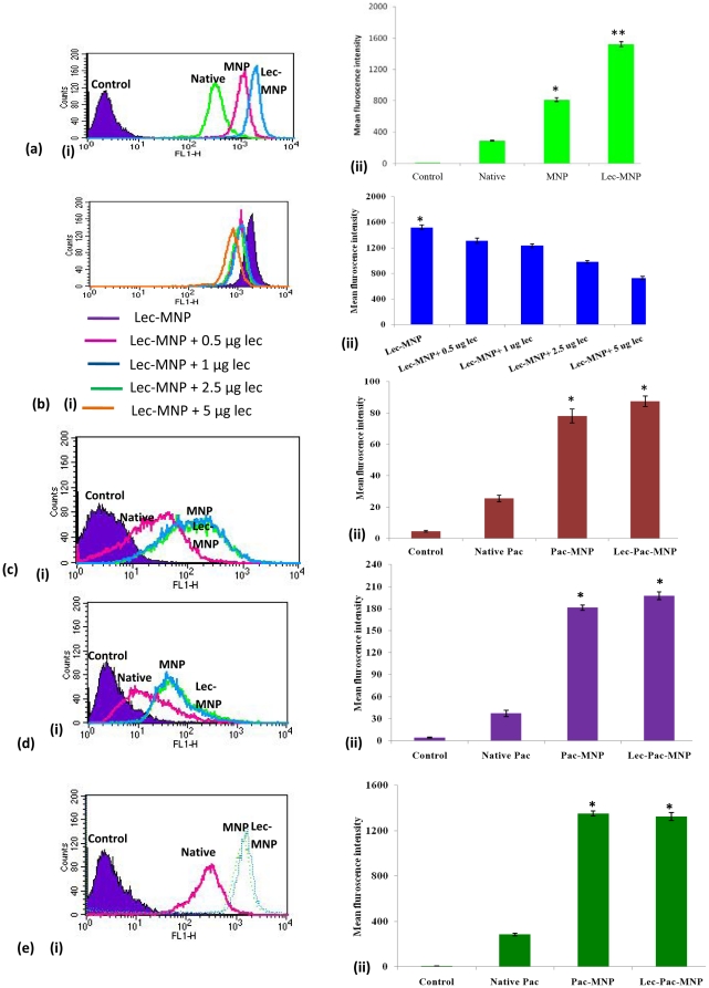
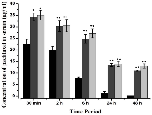
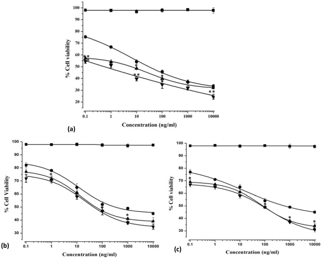
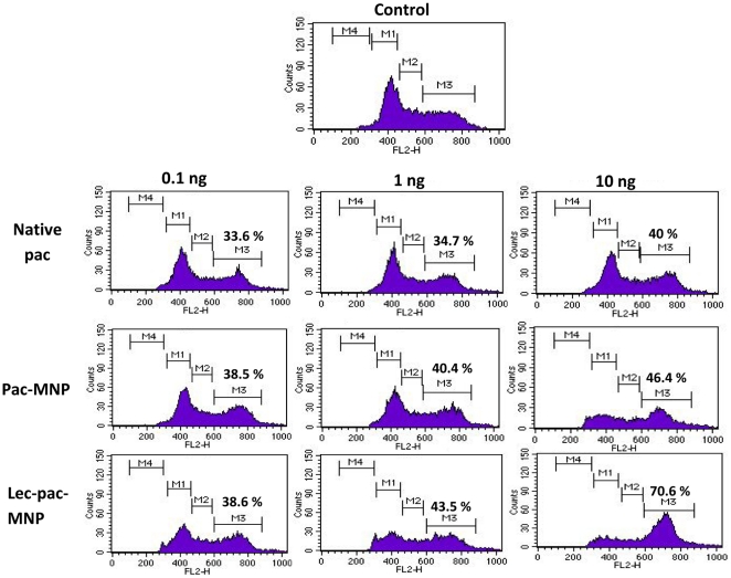
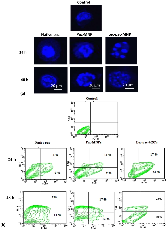
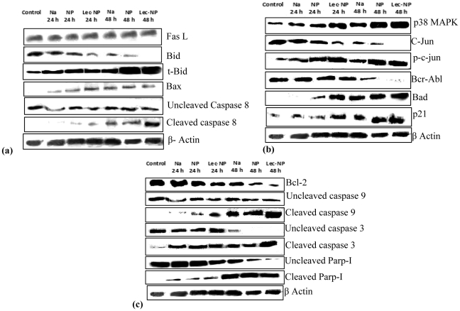
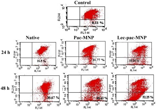
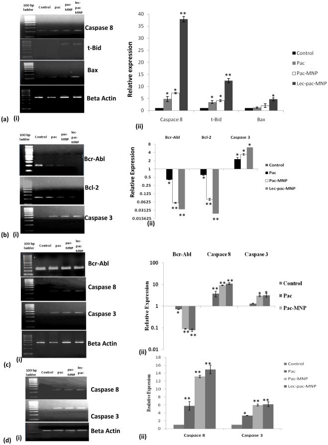
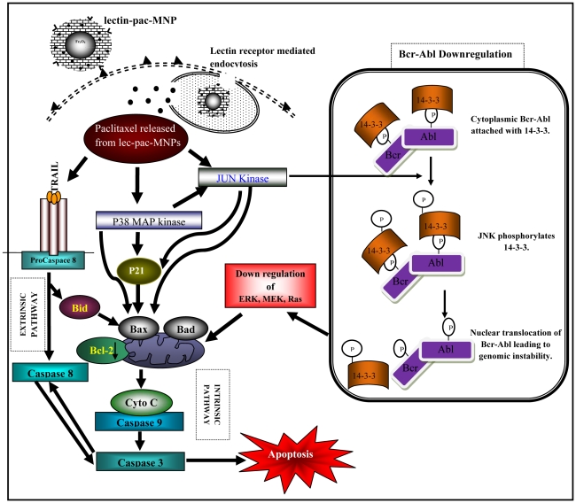
Amongst all leukemias, Bcr-Abl positive chronic myelogenous leukemia (CML) confers resistance to native drug due to multi drug resistance and also resistance to p53 and fas ligand pathways. In the present study, we have investigated the efficacy of microtubule stabilizing paclitaxel loaded magnetic nanoparticles (pac-MNPs) to ascertain its cytotoxic effect on Bcr-Abl positive K562 cells. For active targeted therapy, pac-MNPs were functionalized with lectin glycoprotein which resulted in higher cellular uptake and lower IC(50) value suggesting the efficacy of targeted delivery of paclitaxel. Both pac-MNPs and lectin conjugated pac-MNPs have a prolonged circulation time in serum suggesting increased bioavailability and therapeutics index of paclitaxel in vivo. Further, the molecular mechanism pertaining to pac-induced cytotoxicity was analyzed by studying the involvement of different apoptotic pathway proteins by immunoblotting and quantitative PCR. Our study revealed simultaneous activation of JNK pathway leading to Bcr-Abl instability and the extrinsic apoptotic pathway after pac-MNPs treatment in two Bcr-Abl positive cell lines. In addition, the MRI data suggested the potential application of MNPs as imaging agent. Thus our in vitro and in vivo results strongly suggested the pac-MNPs as a future prospective theranostic tool for leukemia therapy.


Conflict of interest statement

Competing Interests: The authors have declared that no competing interests exist.

Figures

Figure 1
Figure 1. Physicochemical characterization of pac-MNPs.
(A) TEM figure of pac-MNPs. (B) AFM figure of pac-MNPs. (C) DSC curves of (i) native pac, (ii) void MNPs and (iii) pac-MNPs. (D) Release kinetics of paclitaxel from pac-MNPs and lec-pac-MNPs (data as mean ± SEM, n = 3). The insert represents the release of paclitaxel from pac-MNPs and lec-pac-MNPs up to 5th day (data as mean ± SD, n = 3).
Figure 2
Figure 2. MRI study.
(A) MR signal intensity of pac-MNPs in phantom agar gel at various iron concentration, blank phantom agar gels were taken as control. (B) T2 relaxation analysis curves of Pac-MNPs in phantom agar gel at different concentration (data as mean intensity within region of interest (ROI)). (C) MR signal intensity of pac-MNPs and lec-pac-MNPs treated K562 cells in phantom agar gel at various cell concentration, blank phantom agar gels were taken as control. (D) T2 relaxation analysis curves of Pac-MNPs and lec-pac-MNPs treated K562 cells in phantom agar gel at different cell concentration (data as mean intensity within region of interest (ROI)).
Figure 3
Figure 3. Uptake studies.
(A) Uptake analysis of native 6-coumarin, 6-coumarin-MNPs and lec-6-coumarin-MNPs in K562 cells (i) through flow cytometry (ii) Mean fluorescence intensity obtained from flow cytometry. (B) Competitive inhibition assay study for uptake of lec-6-coumarin-MNPs by adding different concentrations of native lectin in K562 cells (i) through flow cytometry (ii) Mean fluorescence intensity obtained from flow cytometry. (C) Uptake analysis of native 6-coumarin, 6-coumarin-MNPs and lec-6-coumarin-MNPs in Kcl22 cells (i) through flow cytometry (ii) Mean fluorescence intensity obtained from flow cytometry. (D) Uptake analysis of native 6-coumarin, 6-coumarin-MNPs and lec-6-coumarin-MNPs in Jurkat cells (i) through flow cytometry (ii) Mean fluorescence intensity obtained from flow cytometry. (E) Uptake analysis of native 6-coumarin, 6-coumarin-MNPs and lec-6-coumarin-MNPs in HEK293 cells (i) through flow cytometry (ii) Mean fluorescence intensity obtained from flow cytometry. Data as mean ± S.E.M., n = 6. (*) p<0.05, (**) p <0.01.
Figure 4
Figure 4. In vivo bioavailability of native pac (▪), pac-MNPs (▪) and lec-pac-MNPs (▪) in a rat model.
The rats were divided in three groups. Equivalent concentration of pac, pac-MNPs and lec-pac-MNPs (20 mg/kg) was administered intravenously. Blood was collected at different time intervals and paclitaxel concentration in serum was determined by RP-HPLC analysis, as described in materials and methods. (**) p<0.01 and (*) p<0.05 pac versus pac-MNPs.
Figure 5
Figure 5. Dose dependent cytotoxicity studies of Void MNP (▪), native pac (•), pac-MNPs (▴) and lec-pac MNPs (∇) in (A) K562 cells, (B) Kcl22 cells and (C) Jurkat cells after 48 h.
The extent of growth inhibition was measured by the MTT assay. The inhibition was calculated with respect to control (cells treated with only medium). Data as mean ± S.E.M., n = 6. (*) p<0.05, drug in solution versus drug in MNPs, (**) p<0.01 drug in solution versus lec-pac-MNPs.
Figure 6
Figure 6. Cell cycle analysis of K562 cells after 24 h of treatment with native pac, pac-MNPs and lec-pac-MNPs at concentration of 0.1 ng, 1 ng and 10 ng/ml.
The figure shows the percentage of cells at G2-M phase of cell cycle.
Figure 7
Figure 7. Apoptosis study in K562 cells.
(A) Morphological analysis of K562 cells after treatment with native pac, pac-MNPs and lec-pac-MNPs (Conc. 10 ng/ml) after 24 and 48 h by confocal microscopy. (B) Induction of apoptosis in K562 cell line by native pac, pac-MNPs and lec-pac-MNPs (Conc. 10 ng/ml) after 24 and 48 h using Annexin V- FITC. Flow cytometry analysis reveals the presence of different populations of cells. Top right: late apoptotic and necrotic cells; bottom left: live cells and bottom right: early apoptotic cells.
Figure 8
Figure 8. Immuno-blot analysis of proteins apoptotic pathway.
(A) extrinsic pathway (B) JNK pathway and (C) intrinsic pathway proteins in K562 cells treated with 10 ng/ml of native pac, pac-MNPs and lec-pac-MNPs (details described in materials and methods.
Figure 9
Figure 9. Changes in mitochondrial membrane potential measured through JC-1 dye after 24 and 48 h of treatment with native pac, pac-MNPs and lec-pac-MNPs (10 ng/ml) in K562 cells.
Figure 10
Figure 10. Gene expression study by real time PCR.
(A) (i) m-RNA expression (ii) quantification of Caspase 8, Bid, and Bax gene by RT-PCR in K562 cell lines. (B) (i) m-RNA expression (ii) quantification of Bcr-Abl, Bcl-2 and caspase 3 gene by RT-PCR in K562 cell lines. (C) (i) m-RNA expression (ii) quantification of Bcr-Abl, Caspase 8 and caspase 3 gene by RT-PCR in Kcl22 cell lines. (D) (i) m-RNA expression (ii) quantification of Caspase 8 and caspase 3 gene by RT-PCR in Jurkat cell lines. (**) p<0.01 and (*) p<0.05 control versus pac, pac-MNPs and lec-pac-MNPs. (Details described in materials and methods).
Figure 11
Figure 11. Schematic diagram showing the receptor mediated endocytosis of lec-pac-MNPs and mechanism of action of paclitaxel after released from the nanoformulation showing coordinated regulation of signaling cascades leading to apoptosis.

Similar articles

Cited by

References

    1. Jacquel A, Herrant M, Legros L, Belhacene N, Luciano F, et al. Imatinib induces mitochondria-dependent apoptosis of the Bcr-Abl-positive K562 cell line and its differentiation toward the erythroid lineage. Faseb J. 2003;17:2160–2162. - PubMed
    1. Terme M, Borg C, Guilhot F, Masurier C, Flament C, et al. BCR/ABL promotes dendritic cell-mediated natural killer cell activation. Cancer Res. 2005;65:6409–6417. - PubMed
    1. Nagata Y, Todokoro K. Requirement of activation of JNK and p38 for environmental stress-induced erythroid differentiation and apoptosis and of inhibition of ERK for apoptosis. Blood. 1999;94:853–863. - PubMed
    1. Jia L, Patwari Y, Kelsey SM, Newland AC. Trail-induced apoptosis in Type I leukemic cells is not enhanced by overexpression of bax. Biochem Biophys Res Commun. 2001;283:1037–1045. - PubMed
    1. Knijn A, Brisdelli F, Ferretti A, Iorio E, Marcheggiani D, et al. Metabolic alterations in K562 cells exposed to taxol and tyrphostin AG957: 1H NMR and biochemical studies. Cell Biol Int. 2005;29:890–897. - PubMed

MeSH terms