

The nuclear pore complex mediates binding of the Mig1 repressor to target promoters
- PMID: 22110603
- PMCID: PMC3215702
- DOI: 10.1371/journal.pone.0027117
The nuclear pore complex mediates binding of the Mig1 repressor to target promoters
Abstract
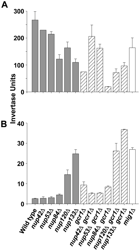
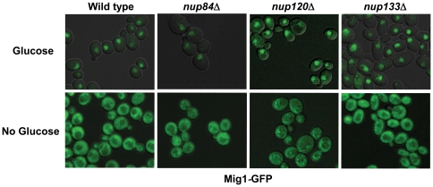
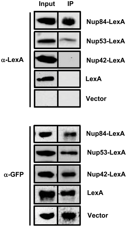
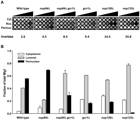
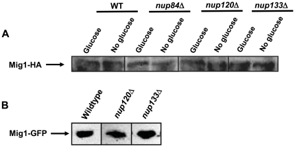
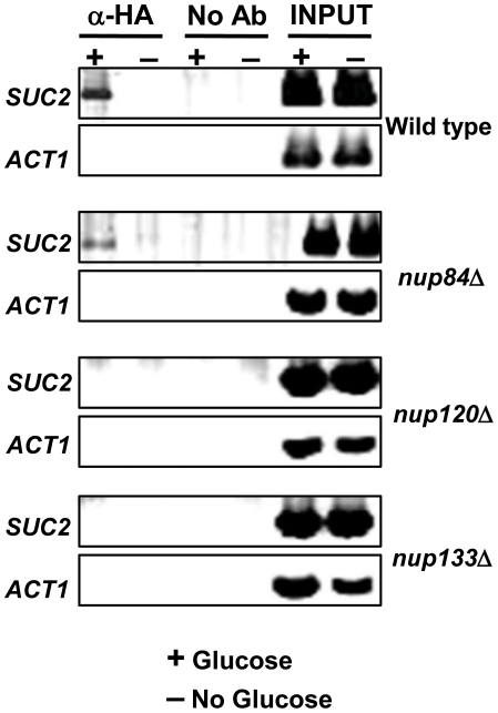
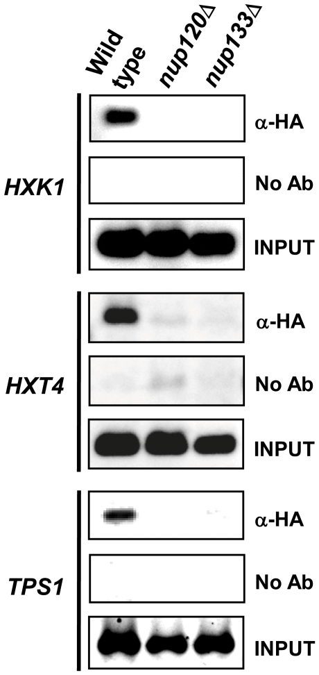
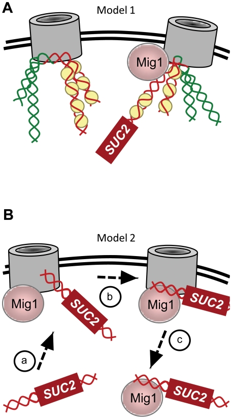
All eukaryotic cells alter their transcriptional program in response to the sugar glucose. In Saccharomyces cerevisiae, the best-studied downstream effector of this response is the glucose-regulated repressor Mig1. We show here that nuclear pore complexes also contribute to glucose-regulated gene expression. NPCs participate in glucose-responsive repression by physically interacting with Mig1 and mediating its function independently of nucleocytoplasmic transport. Surprisingly, despite its abundant presence in the nucleus of glucose-grown nup120Δ or nup133Δ cells, Mig1 has lost its ability to interact with target promoters. The glucose repression defect in the absence of these nuclear pore components therefore appears to result from the failure of Mig1 to access its consensus recognition sites in genomic DNA. We propose that the NPC contributes to both repression and activation at the level of transcription.
Conflict of interest statement
Figures
Similar articles
-
Hxk2 regulates the phosphorylation state of Mig1 and therefore its nucleocytoplasmic distribution.J Biol Chem. 2007 Feb 16;282(7):4485-4493. doi: 10.1074/jbc.M606854200. Epub 2006 Dec 18. J Biol Chem. 2007. PMID: 17178716
-
Mig1 localization exhibits biphasic behavior which is controlled by both metabolic and regulatory roles of the sugar kinases.Mol Genet Genomics. 2020 Nov;295(6):1489-1500. doi: 10.1007/s00438-020-01715-4. Epub 2020 Sep 19. Mol Genet Genomics. 2020. PMID: 32948893 Free PMC article.
-
The glucose-regulated nuclear localization of hexokinase 2 in Saccharomyces cerevisiae is Mig1-dependent.J Biol Chem. 2004 Apr 2;279(14):14440-6. doi: 10.1074/jbc.M313431200. Epub 2004 Jan 8. J Biol Chem. 2004. PMID: 14715653
-
The new nucleoporin: regulator of transcriptional repression and beyond.Nucleus. 2012 Nov-Dec;3(6):508-15. doi: 10.4161/nucl.22427. Epub 2012 Oct 9. Nucleus. 2012. PMID: 23047597 Free PMC article.
-
Glucose control in Saccharomyces cerevisiae: the role of Mig1 in metabolic functions.Microbiology (Reading). 1998 Jan;144 ( Pt 1):13-24. doi: 10.1099/00221287-144-1-13. Microbiology (Reading). 1998. PMID: 9467897 Review. No abstract available.
Cited by
-
Genetic interaction network has a very limited impact on the evolutionary trajectories in continuous culture-grown populations of yeast.BMC Ecol Evol. 2021 May 26;21(1):99. doi: 10.1186/s12862-021-01830-9. BMC Ecol Evol. 2021. PMID: 34039270 Free PMC article.
-
Nuclear pore proteins and the control of genome functions.Genes Dev. 2015 Feb 15;29(4):337-49. doi: 10.1101/gad.256495.114. Genes Dev. 2015. PMID: 25691464 Free PMC article. Review.
-
Sumoylation and transcription regulation at nuclear pores.Chromosoma. 2015 Mar;124(1):45-56. doi: 10.1007/s00412-014-0481-x. Epub 2014 Aug 30. Chromosoma. 2015. PMID: 25171917 Free PMC article. Review.
-
Nuclear routing networks span between nuclear pore complexes and genomic DNA to guide nucleoplasmic trafficking of biomolecules.J Fertili In Vitro. 2012 Oct 19;2(4):112. doi: 10.4172/2165-7491.1000112. J Fertili In Vitro. 2012. PMID: 23275893 Free PMC article.
-
A negative feedback loop at the nuclear periphery regulates GAL gene expression.Mol Biol Cell. 2012 Apr;23(7):1367-75. doi: 10.1091/mbc.E11-06-0547. Epub 2012 Feb 9. Mol Biol Cell. 2012. PMID: 22323286 Free PMC article.
References
-
- Towler MC, Hardie DG. AMP-activated protein kinase in metabolic control and insulin signaling. Circ Res. 2007;100:328–341. - PubMed
-
- Winder WW, Hardie DG. AMP-activated protein kinase, a metabolic master switch: possible roles in type 2 diabetes. Am J Physiol. 1999;277:E1–10. - PubMed
-
- Hong YH, Varanasi US, Yang W, Leff T. AMP-activated protein kinase regulates HNF4alpha transcriptional activity by inhibiting dimer formation and decreasing protein stability. J Biol Chem. 2003;278:27495–27501. - PubMed
-
- Leff T. AMP-activated protein kinase regulates gene expression by direct phosphorylation of nuclear proteins. Biochem Soc Trans. 2003;31:224–227. - PubMed
Publication types
MeSH terms
Substances
Grants and funding
LinkOut - more resources
Full Text Sources
Molecular Biology Databases
