. 2011;6(11):e27246.
doi: 10.1371/journal.pone.0027246. Epub 2011 Nov 3.

Fatal S. aureus hemorrhagic pneumonia: genetic analysis of a unique clinical isolate producing both PVL and TSST-1

Affiliations

Fatal S. aureus hemorrhagic pneumonia: genetic analysis of a unique clinical isolate producing both PVL and TSST-1

Zhi Li et al. PLoS One. 2011.

Abstract

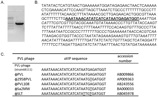
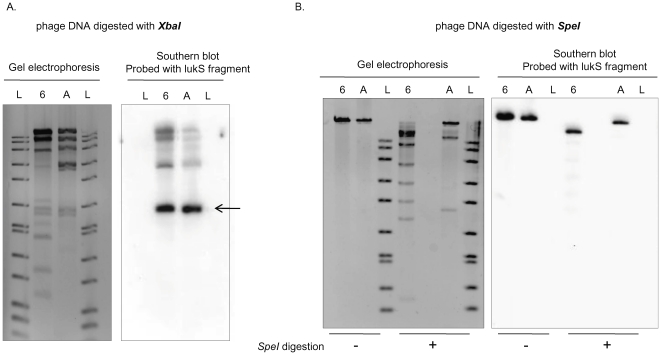
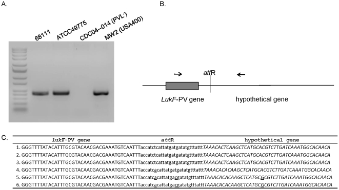
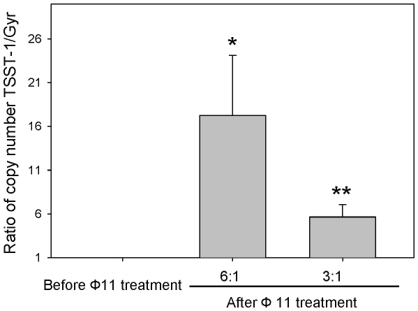
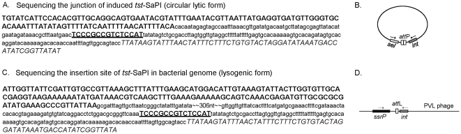
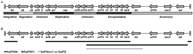
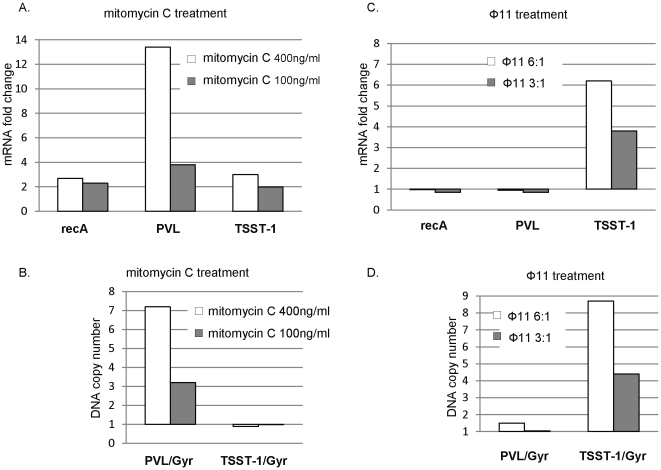
In 2008, an unusual strain of methicillin-sensitive Staphylococcus aureus (MSSA68111), producing both Panton-Valentine leukocidin (PVL) and toxic shock syndrome toxin-1 (TSST-1), was isolated from a fatal case of necrotizing pneumonia. Because PVL/TSST-1 co-production in S. aureus is rare, we characterized the molecular organization of these toxin genes in strain 68111. MSSA68111 carries the PVL genes within a novel temperate prophage we call ФPVLv68111 that is most similar, though not identical, to phage ФPVL--a phage type that is relatively rare worldwide. The TSST-1 gene (tst) in MSSA68111 is carried on a unique staphylococcal pathogenicity island (SaPI) we call SaPI68111. Features of SaPI68111 suggest it likely arose through multiple major recombination events with other known SaPIs. Both ФPVLv68111 and SaPI68111 are fully mobilizable and therefore transmissible to other strains. Taken together, these findings suggest that hypervirulent S. aureus have the potential to emerge worldwide.


Conflict of interest statement

Competing Interests: The authors have declared that no competing interests exist.

Figures

Figure 1
Figure 1. Alpha- and delta-hemolysin activities of S. aureus strain 68111.
A plate-based screening method that capitalizes on beta-hemolysin-mediated effects on alpha- and delta-hemolysin activities was used to examine extracellular production of these toxins in MSSA68111. For this, beta-hemolysin-producing S. aureus strain RN4220 was streaked vertically on sheep blood agar. MSSA68111 (top) and two laboratory control strains (α-hemolysinlow/δ-hemolysin- [middle] and α-hemolysin+/δ-hemolysin+ [bottom]) were then streaked perpendicularly to RN4220. Hemolysis was assessed after 24 hrs incubation at 37°C (no cold shock applied). At the junction between the test strains and RN4220, alpha-hemolysin activity is inhibited (arrowhead); the presence of delta-hemolysin is observed as an enhanced area of hemolysis (arrows).
Figure 2
Figure 2. DNA sequence of the attP junction of MSSA68111 PVL phage.
(A.) PCR was performed with an inverse primer set using mitomycin C-elicited phage genomic DNA from MSSA68111 as a template. PCR products were separated by agarose gel electrophoresis and stained with ethidium bromide. (B.) Nucleotide sequence of the 68111 PCR product was determined. The sequence originating from PVL phage integrase is printed in upper-case letters, and that derived from lukF-PV gene is shown in italic letters, and twenty-nine base pairs of the core attP sequence are bolded and underlined. (C.) This attP sequence of MSSA68111 PVL phage was compared to that from other published PVL-carrying phages. Nucleotide variations were underlined.
Figure 3
Figure 3. Southern blot analysis of PVL phage genomic DNA from strain 68111 and strain ATCC49775.
Phage DNA from MSSA68111 (Lanes designated “6”) and from strain ATCC49775 (Lanes “A”) was digested with either XbaI (A.) or SpeI (B.). In both A and B, the panels on the left are photographs of ethidium bromide-stained gels; the panels on the right are Southern blots hybridized with the lukS-PV probe. 1 kb DNA ladders (Lanes “L”) are shown. The lukS-PV probe hybridized to identical 2 kb fragments in XbaI -digested phage DNA from both strains (A, arrow) whereas SpeI-digestion produced different sized lukS-PV fragments (B.).
Figure 4
Figure 4. DNA sequences of integration site of PVL phage in bacterial genome.
(A.) PCR was performed with primers, as shown in (B.) using bacterial genomic DNA as templates. PCR products were separated by agarose gel electrophoresis and stained with ethidium bromide. (C.) The sequence of PCR product from MSSA68111 was determined and is shown relative to other strains carrying PVL phage. The sequence originating from the phage PVL genomic DNA including lukF-PV gene is printed in upper-case letters, and that derived from the bacterial chromosome, a conserved hypothetical protein, is shown in italic letters. Twenty-nine base pairs of core attR sequence are bolded and underlined. Nucleotide variations are underlined. 1.ФPVLv68111; 2. ΦPVL (strain ATCC49775 [25]); 3. ΦPVL108 (Accession: AB243556); 4. Φ2958 PVL (Accession:AP009363); 5. ΦSLT (Accession: CP000255); 6. ΦSa2 MW (Accession: BA000033).
Figure 5
Figure 5. Induction (excision/replication) of tst-SaPI elements by phage11.
Following superinfection with phage 11 as described in Materials and Methods, total genomic DNA was purified and analyzed for tst and gyr genes by real-time PCR. Relative copy numbers of tst versus gyr are presented. Data are means±SD (n = 4); p*<0.05; p**<0.01 vs control (before Ф11 treatment) (paired t-test).
Figure 6
Figure 6. Sequence analyses of tst-carrying SaPI in MSSA68111.
(A.) Junctional sequence of circulated SaPI (lytic form) following helper phage 11 induction. Sel coding region is bolded; integrase of tst-SaPI is shown in italic letter and 15 base pairs of core attP are bolded and underlined. Other intergenic regions are presented in lower-case font. A schematic illustration of the PCR strategy used to amplify this junctional region is shown in (B.). (C.) Sequence of integration site of tst-SaPI (lysogenic form) in bacterial genome. SsrP coding region is bolded, integrase of tst-SaPI is shown in italic letter and 15 base pairs of core attL are bolded and underlined. Other intergenic regions are presented in lower-case font. A schematic illustration of the PCR strategy used to amplify the integration site of tst-SaPI is shown in (D.).
Figure 7
Figure 7. Illustrative representation of pathogencity island 68111 (SaPI68111).
Arrows represent the location and orientation of open reading frames (OFRs), which were determined using the NCBI-GLIMMER program and by the relative position of ORFs within the island and/or similarity of the predicted gene products. (A.) An illustrative diagram of the SaPI68111 genome with different functional annotations presented at the bottom of the figure. (B.) A summary diagram of the homology regions between SaPI68111 and other close-related SaPIs. SaPIM4 (thick line), SaPIM1 (triple lines) and SaPIbov1 or SaPI2 (thin line).
Figure 8
Figure 8. Amino acid alignment of predicted stl from MSSA68111 and hypothetical phage repressor protein SH2101 from Staphylococcus haemolyticus (Accession: YP_254016.1).
The protein sequences were aligned by using the CLUSTAL alignment program. The Helix-turn-helix XRE-domain is highlighted in the black box. The asterisk denotes a position at which the two sequences have the same amino acid. Dots indicate the degree of homology when there is not complete sequence conservation.
Figure 9
Figure 9. Comparative study of phage induction versus gene transcription in response to mitomycin C or phage 11 treatment.
(A. and B.) Bacteria were treated with mitomycin C (400 ng/ml or 100 ng/ml) for 1 hr. (C. and D.) Freshly prepared phage 11 was added for 1 hr. One half of samples were used for RNA preparation followed by a quantitative RT-PCR analysis. Fold changes in gene expression of recA, PVL and TSST-1 were normalized to the internal control gene gyrase (Gyr) and relative to no treatment control sample. The other half of samples were used for total genomic DNA preparation. Relative copy number of TSST-1 or PVL gene versus Gyr were calculated as described in Materials and Methods. Shown is one representative out of three independent experiments, all of which gave comparable results.

References

    1. Archer GL. Staphylococcus aureus: a well-armed pathogen. Clin Infect Dis. 1998;26:1179–1181. - PubMed
    1. Lowy FD. Staphylococcus aureus infections. N Engl J Med. 1998;339:520–532. 10.1056/NEJM199808203390806 [doi] - PubMed
    1. Kaneko J, Kamio Y. Bacterial two-component and hetero-heptameric pore-forming cytolytic toxins: structures, pore-forming mechanism, and organization of the genes. Biosci Biotechnol Biochem. 2004;68:981–1003. - PubMed
    1. Gillet Y, Issartel B, Vanhems P, Fournet JC, Lina G, et al. Association between Staphylococcus aureus strains carrying gene for Panton-Valentine leukocidin and highly lethal necrotising pneumonia in young immunocompetent patients. Lancet. 2002;359:753–759. S0140-6736(02)07877-7 [pii];10.1016/S0140-6736(02)07877-7 [doi] - PubMed
    1. See RH, Kum WW, Chang AH, Goh SH, Chow AW. Induction of tumor necrosis factor and interleukin-1 by purified staphylococcal toxic shock syndrome toxin 1 requires the presence of both monocytes and T lymphocytes. Infect Immun. 1992;60:2612–2618. - PMC - PubMed

Publication types

MeSH terms

LinkOut - more resources