. 2011;6(11):e27352.
doi: 10.1371/journal.pone.0027352. Epub 2011 Nov 14.

Mitochondrial-associated cell death mechanisms are reset to an embryonic-like state in aged donor-derived iPS cells harboring chromosomal aberrations

Affiliations

Mitochondrial-associated cell death mechanisms are reset to an embryonic-like state in aged donor-derived iPS cells harboring chromosomal aberrations

Alessandro Prigione et al. PLoS One. 2011.

Abstract

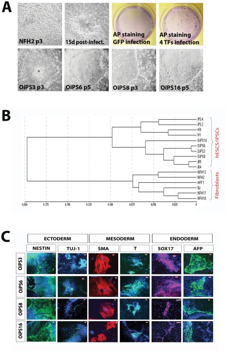
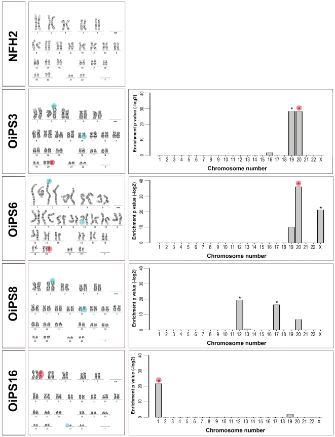
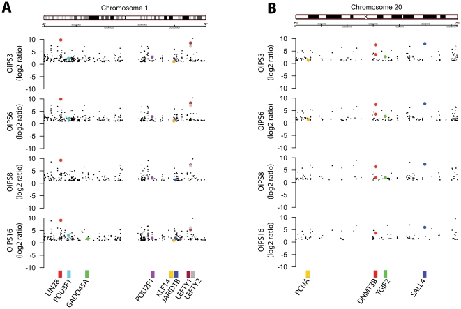
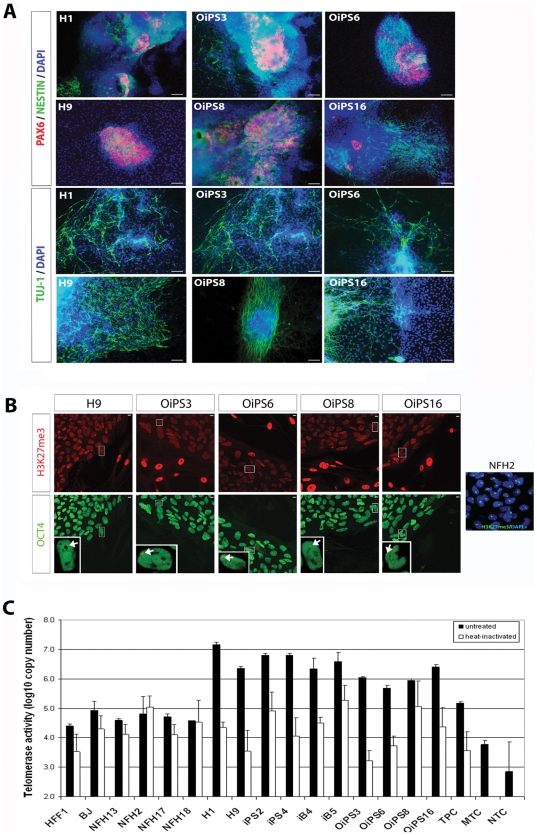
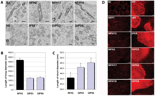
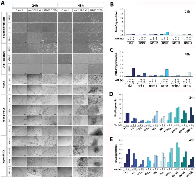
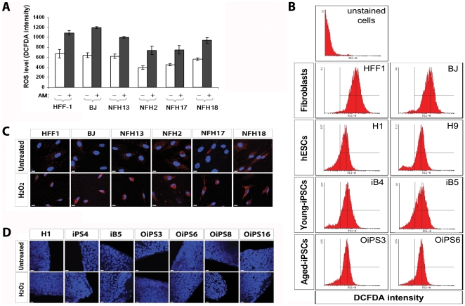
Somatic cells reprogrammed into induced pluripotent stem cells (iPSCs) acquire features of human embryonic stem cells (hESCs) and thus represent a promising source for cellular therapy of debilitating diseases, such as age-related disorders. However, reprogrammed cell lines have been found to harbor various genomic alterations. In addition, we recently discovered that the mitochondrial DNA of human fibroblasts also undergoes random mutational events upon reprogramming. Aged somatic cells might possess high susceptibility to nuclear and mitochondrial genome instability. Hence, concerns over the oncogenic potential of reprogrammed cells due to the lack of genomic integrity may hinder the applicability of iPSC-based therapies for age-associated conditions. Here, we investigated whether aged reprogrammed cells harboring chromosomal abnormalities show resistance to apoptotic cell death or mitochondrial-associated oxidative stress, both hallmarks of cancer transformation. Four iPSC lines were generated from dermal fibroblasts derived from an 84-year-old woman, representing the oldest human donor so far reprogrammed to pluripotency. Despite the presence of karyotype aberrations, all aged-iPSCs were able to differentiate into neurons, re-establish telomerase activity, and reconfigure mitochondrial ultra-structure and functionality to a hESC-like state. Importantly, aged-iPSCs exhibited high sensitivity to drug-induced apoptosis and low levels of oxidative stress and DNA damage, in a similar fashion as iPSCs derived from young donors and hESCs. Thus, the occurrence of chromosomal abnormalities within aged reprogrammed cells might not be sufficient to over-ride the cellular surveillance machinery and induce malignant transformation through the alteration of mitochondrial-associated cell death. Taken together, we unveiled that cellular reprogramming is capable of reversing aging-related features in somatic cells from a very old subject, despite the presence of genomic alterations. Nevertheless, we believe it will be essential to develop reprogramming protocols capable of safeguarding the integrity of the genome of aged somatic cells, before employing iPSC-based therapy for age-associated disorders.


Conflict of interest statement

Competing Interests: The authors have declared that no competing interests exist.

Figures

Figure 1
Figure 1. Derivation and characterization of four iPSC lines from an 84-year-old woman.
(A) NFH2 dermal fibroblasts were obtained from an 84-year-old woman and transduced using retroviruses containing the four transcription factors (TFs) OCT4, KLF4, SOX2 and c-MYC. Four weeks later, hESC-like colonies were stained for alkaline phosphatase (AP) activity then manually picked for further characterization. Four distinct iPSC lines were established: OiPS3, OiPS6, OiPS8, and OiPS16. (B) Hierarchical clustering of all fibroblast cells and pluripotent stem cells. NFH2-derived iPSC lines acquired a pluripotent stem cell-like transcriptional signature and clustered far apart from the parental NFH2 fibroblasts and from other fibroblast cells. (C) Embryoid body (EB)-based in vitro differentiation of the four NFH2-derived iPSC lines. All differentiated NFH2-iPSC lines expressed marker proteins specific to the three germ layers: ectoderm (NESTIN and TUJ-1), mesoderm (SMOOTH MUSCLE ACTIN (SMA) and BRACHYURY (T)), and endoderm (SOX17 and ALPHA FETO PROTEIN (AFP)). Scale bars, 20 µm.
Figure 2
Figure 2. Chromosomal aberrations in aged reprogrammed cells.
Left panel, karyotype analysis of the parental NFH2 fibroblast cells and NFH2-derived iPSC lines. NFH2 was karyotypically normal, while all NFH2-iPSC lines exhibited chromosomal aberrations (red circles and light blue circles). Right panel, transcriptional profiling-based analysis of chromosomal integrity. Expression values of single genes in NFH2-iPSC lines were compared to the parental NFH2 fibroblasts and considered over-expressed when fold-change>2 and Bonferroni corrected p-values <10−4. The analysis confirmed significant enrichment in all cases in which karyotyping showed trisomic transformation (red circles). Additional significant enrichments could be observed in NFH2-iPSCs in chromosomes that appeared normal upon karyotyping. Chromosomal translocations (10 to 2) in OiPS3, OiPS6, OiPS8 and chromosomal 1 loss in OiPS16 could not be confirmed by gene expression-based chromosomal enrichment analysis (light blue circles).
Figure 3
Figure 3. Enrichment of pluripotency and tumorigenicity-associated genes within chromosomes 1 and 20 in aged-iPSCs.
(A) Moving average plot analysis of chromosome 1. Expression values in NFH2-iPSC lines were compared to the parental NFH2 fibroblasts and considered over-expressed or under-expressed when fold-change>2. Each dot represents a probeset encompassing an individual gene. Highlighted in different colors are probeset for genes whose function has been associated with pluripotency, reprogramming, proliferation, or tumorigenicity. (B) Moving average plot analysis of chromosome 20. Moving average plots were produced using R/Bioconductor package GenomeGraphs.
Figure 4
Figure 4. Neuronal differentiation, X-chromosome inactivation, and telomerase activity in aged donor-derived iPSCs.
(A) Neuronal cells were derived by the combined treatment of transforming growth factor β(TGFβ) receptor inhibitor and MEK1/2 inhibitor. All aged-iPSC lines generated neuronal-like cells expressing PAX6, NESTIN, and TUJ-1, in a similar fashion to the hESC lines H1 and H9. Scale bars, 10 µm. (B) X-chromosome inactivation (XCI) was assayed by H3K27me3 immunostaining. OCT4 staining was used to distinguish undifferentiated pluripotent stem cells from MEFs. NFH2 fibroblasts clearly exhibited inactive X-chromosomes (Xi), while variable XCI status was observed in both H9 and NFH2-derived iPSC lines. Single female pluripotent cells expressing Xi are indicated by the white arrows in the bottom inserts. Scale bars, 10 µm. (C) Telomerase activity was low in fibroblasts from neonates (HFF1 and BJ) and from adult individuals of various ages (NFH13-55y, NFH2-84y, NFH17-80y, and NFH18-80y). The level of activity was similarly elevated in all pluripotent stem cells, including hESCs (H1 and H9), HFF1-iPSCs (iPS2 and iPS4), BJ-iPSCs (iB4 and iB5) and NFH2-iPSCs (OiPS3, OiPS6, OiPS8, and OiPS16). Heat inactivation was performed to show the different enzymatic activity between the active and inactive state. TPC, telomerase positive extract control (100 ng of positive control cell protein extract provided by the kit); MTC, minus telomerase control (cell sample substituted by CHAPS Lysis Buffer); NTC, no template Control (cell sample substituted by water). Error bars indicate standard deviation.
Figure 5
Figure 5. iPSCs from an 84-year-old woman acquired embryonic-like mitochondrial ultra-structure and functionality.
(A) Transmission electron microscopy (TEM) analysis showing mitochondrial ultra-structural morphology in fibroblasts and in pluripotent stem cells. HFF1, neonatal fibroblasts; NFH2, dermal fibroblasts from an 84-year-old woman; NFH17 from an 80-year-old man; NFH18 from an 80-year-old woman; H9, female hESC line; iPS4, iPSC line derived from HFF1 fibroblasts; OiPS3 and OiPS6, iPSC lines generated from NFH2 fibroblasts. Scale bars, 500 nm. (B–C) Measurement of long and short mitochondrial diameters in NFH2 fibroblasts and in two NFH2-derived iPSC lines (OiPS3 and OiPS6). 50 mitochondria were measured for each sample using the EMMENU4 software (Fastscan, TVIPS). Upon reprogramming the long diameter becomes shorter while the short diameter increases, suggesting that the organelles tend to acquire a round-like shape. Error bars indicate the standard error of means (SEM). (D) Mitochondrial membrane potential (MMP) was assessed employing the fluorescent dye TMRE in live cells to obtain a relative measurement of mitochondria functionality in terms of energy coupling. All human fibroblasts exhibited low basal MMP. The fluorescent intensity appeared increased in undifferentiated hESCs and iPSCs, while differentiated cells at the borders of pluripotent stem cell colonies showed reduction of basal MMP signal. Thus, the mitochondrial functionality of aged iPSCs appeared similar of that of young iPSCs and hESCs. The presence of mitochondrial hyper-polarization, suggestive of reduced ATP consumption, in all pluripotent stem cells is in agreement with the distinctive metabolic features of undifferentiated stem cells which rely more on glycolytic rather than oxidative-based energy generation. Scale bars, 50 µm.
Figure 6
Figure 6. Sensitivity to drug-induced apoptosis in young and aged fibroblasts, hESCs, young-iPSCs, and aged-iPSCs.
(A) Apoptotic cell death was measured both at the basal level and upon the treatment with two different doses of actinomycin D (AM) (10−9 M and 10−7 M). Pictures were taken after 24 h and 48 h. Scale bars, 100 µm. (B) Induction of apoptosis after 24 h AM treatment in young and aged-derived fibroblasts. (C) Apoptosis levels after 48 h AM treatment in young and aged-derived fibroblasts. (D) Apoptotic cell death at 24 h in hESCs, young-iPSCs (iPS2, iPS4, iB4, iB5) and aged-iPSCs (OiPS3, OiPS6, OiPS8, OiPS16). (E) Induction of apoptosis after 48 h AM treatment in pluripotent stem cells.
Figure 7
Figure 7. ROS and DNA damage in aged donor-derived iPSCs.
(A) Production of ROS in fibroblast cells was measured by FACS using DCF-DA at basal level and after 24 h treatment with high doses actinomycin D (AM) (10−6 M). Analysis was conducted in quadruplicate and reported as fluorescence intensity. Error bars indicate standard deviation. (B) Representative DCF-DA-based FACS measurements of basal ROS generation in two fibroblasts (HFF1 and BJ), two hESC lines (H1 and H9), two young donor-derived iPSC lines (iB4 and iB5), and two aged donor-derived iPSC lines (OiPS3 and OiPS6). (C) Oxidative damage to nuclear and mitochondrial DNA within fibroblast cells was assessed by 8-hydroxy-2′-deoxyguanosine (8OHdG) staining. Treatment with 500 µM H2O2 for two hours was employed to trigger oxidative-mediated DNA damage. Scale bars, 10 µm. (D) Embryonic and somatic-derived pluripotent stem cells were stained with an antibody against 8OHdG to detect nuclear or mitochondrial DNA lesions induced by oxidative stress. The analysis was conducted in standard growth conditions and after a 2 h exposure to 500 µM H2O2. Scale bars, 10 µm.

References

    1. Takahashi K, Tanabe K, Ohnuki M, Narita M, Ichisaka T, et al. Induction of pluripotent stem cells from adult human fibroblasts by defined factors. Cell. 2007;131:861–872. - PubMed
    1. Yu J, Vodyanik MA, Smuga-Otto K, Antosiewicz-Bourget J, Frane JL, et al. Induced pluripotent stem cell lines derived from human somatic cells. Science. 2007;318:1917–1920. - PubMed
    1. Jozefczuk J, Prigione A, Chavez L, Adjaye J. Comparative Analysis of Human Embryonic Stem Cell and Induced Pluripotent Stem Cell-Derived Hepatocyte-Like Cells Reveals Current Drawbacks and Possible Strategies for Improved Differentiation. Stem Cells Dev. 2011. - PubMed
    1. Kiskinis E, Eggan K. Progress toward the clinical application of patient-specific pluripotent stem cells. J Clin Invest. 2010;120:51–59. - PMC - PubMed
    1. Laurent LC, Ulitsky I, Slavin I, Tran H, Schork A, et al. Dynamic changes in the copy number of pluripotency and cell proliferation genes in human ESCs and iPSCs during reprogramming and time in culture. Cell Stem Cell. 2011;8:106–118. - PMC - PubMed

Publication types