. 2011;6(11):e27485.
doi: 10.1371/journal.pone.0027485. Epub 2011 Nov 14.

Autophagy in human embryonic stem cells

Affiliations

Autophagy in human embryonic stem cells

Thien Tra et al. PLoS One. 2011.

Abstract

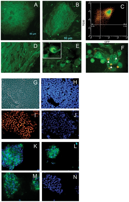
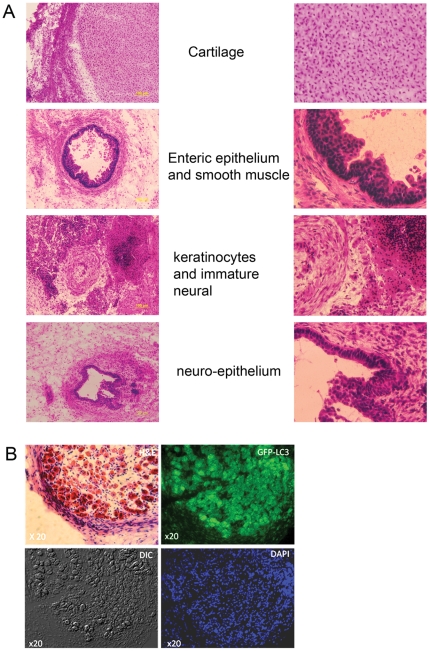
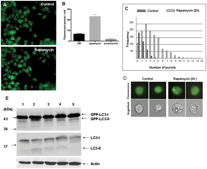
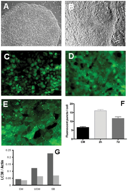
Autophagy (macroautophagy) is a degradative process that involves the sequestration of cytosolic material including organelles into double membrane vesicles termed autophagosomes for delivery to the lysosome. Autophagy is essential for preimplantation development of mouse embryos and cavitation of embryoid bodies. The precise roles of autophagy during early human embryonic development, remain however largely uncharacterized. Since human embryonic stem cells constitute a unique model system to study early human embryogenesis we investigated the occurrence of autophagy in human embryonic stem cells. We have, using lentiviral transduction, established multiple human embryonic stem cell lines that stably express GFP-LC3, a fluorescent marker for the autophagosome. Each cell line displays both a normal karyotype and pluripotency as indicated by the presence of cell types representative of the three germlayers in derived teratomas. GFP expression and labelling of autophagosomes is retained after differentiation. Baseline levels of autophagy detected in cultured undifferentiated hESC were increased or decreased in the presence of rapamycin and wortmannin, respectively. Interestingly, autophagy was upregulated in hESCs induced to undergo differentiation by treatment with type I TGF-beta receptor inhibitor SB431542 or removal of MEF secreted maintenance factors. In conclusion we have established hESCs capable of reporting macroautophagy and identify a novel link between autophagy and early differentiation events in hESC.


Conflict of interest statement

Competing Interests: The authors have declared that no competing interests exist.

Figures

Figure 1
Figure 1. HES3 LC3-GFP cells are pluripotent.
The majority of cells in an HES3-GFP-LC3 (A) or HES4-GFP-LC3 (B) colony express green fluorescence distributed evenly throughout the cytosol. Flow cytometric analysis of HES3-GFP-LC3 shows greater than 85% of the cells express GFP and the pluripotency marker TG30 (C). GFP-LC3 transduced hESC often show 1–2 bright fluorescent puncta (D) and the appearance of typical autophagosomes (E and insert). A subset of autophagosomes co-localise, as indicated by arrow heads, with the lysosomal membrane marker LAMP-1 (F). GFP-LC3 transduced hESC are small and densely packed when viewed under transmitted light (G) and show a high nuclear to cytoplasm ratio (H, cells stained for DNA with Hoechst). HES3-GFP-LC3 cells express the pluripotency markers Oct4 (I) and corresponding DAPI stained nuclei (J), TRA-181 (K), TRA-160 (L) and TG30 (M). No immunostaining was observed in isotype control stained cells (N).
Figure 2
Figure 2. Teratoma sections derived from HES3 LC3-GFP cells and persistence of LC3-GFP transgene expression.
(A), Representative sections of a 6 week old teratoma derived from HES3-LC3-GFP cells showed cell types representative of the three germlayers with each section shown magnified at right. (B), GFP fluorescence persists in teratomas of HES3-LC3-GFP cells. Representative images showing haemtoxylin/eosin staining and GFP fluorescence of two immediately adjacent serial sections of a HES3-LC3-GFP teratoma are shown. Brightfield and DAPI staining images corresponding to the GFP-fluorescence panel are shown. Arrow heads highlight autophagosomes in themagnified section (inset).
Figure 3
Figure 3. Regulation of autophagy in HES3 LC3-GFP cells.
The number of fluorescent puncta observed by fluorescence microscopy in HES3 LC3-GFP cells under control conditions increased after incubation with rapamycin (200 nM) for 2 h (A). Numbers of fluorescent puncta in cells were determined in control cells and after treatment with rapamycin (200 nM), or wortmannin (150 nM) for 2 h (B). Using fluorescence microscopy over 100 cells were observed in 3 independent experiments. Cells with and without treatment with rapamycin (200 nM) for 2 h analysed by imaging flow cytometry (C). Numbers of fluorescent puncta in 10,000 individual cells from each sample were determined. Representative bright-field and fluorescence emission images for two individual cells from each sample are shown. The fluorescent puncta inside each cell are clearly visible (D). Cell lysates were prepared, subjected to SDS-PAGE and blots probed with a monoclonal antibody against LC3 (top and middle) or actin (lower panel) (E). Conditioned medium (CM) lane 1; CM + rapamycin for 2 h (200 nM), lane 2; unconditioned medium 3 days, lane 3; CM + SB431542 for 3 h (10 µM), lane 4; CM + wortmannin for 3 h (150 nM), lane 5.
Figure 4
Figure 4. Spontaneous and induced differentiation promotes autophagy in HES3 LC3-GFP cells.
Cells were maintained in either conditioned medium (CM, panels A, C) or unconditioned medium (B, D). Bright field images of an individual colony at low magnification (A, B) and fluorescence images are shown. Cells maintained in CM supplemented with SB431542 (10 µM) were imaged after 3 hours incubation (E). Fluorescent puncta were counted in cells for three independent experiments after h and 7 days of incubation in the presence of SB431542 (F). Analysis of western blot data (see Fig 3) was used to calculate the GFP-LC3II/Actin and LC3II/Actin ratios after 3 day culture in unconditioned medium (UCM) and SB431542 treatment for 3 hours (G).

References

    1. Pera MF, Trounson AO. Human embryonic stem cells: prospects for development. Development. 2004;131:5515–5525. - PubMed
    1. Ichimura Y, Kirisako T, Takao T, Satomi Y, Shimonishi Y, et al. A ubiquitin-like system mediates protein lipidation. Nature. 2000;408:488–492. - PubMed
    1. Kabeya Y, Mizushima N, Ueno T, Yamamoto A, Kirisako T, et al. LC3, a mammalian homologue of yeast Apg8p, is localized in autophagosome membranes after processing. EMBO J. 2000;19:5720–5728. - PMC - PubMed
    1. Cullinane M, Gong L, Li X, Lazar-Adler N, Tra T, et al. Stimulation of autophagy suppresses the intracellular survival of Burkholderia pseudomallei in mammalian cell lines. Autophagy. 2008;4:744–753. - PubMed
    1. Klionsky DJ, Cuervo AM, Seglen PO. Methods for monitoring autophagy from yeast to human. Autophagy. 2007;3:181–206. - PubMed

Publication types