. 2011;6(11):e27486.
doi: 10.1371/journal.pone.0027486. Epub 2011 Nov 16.

A unique role of GATA1s in Down syndrome acute megakaryocytic leukemia biology and therapy

Affiliations

A unique role of GATA1s in Down syndrome acute megakaryocytic leukemia biology and therapy

Ana C Xavier et al. PLoS One. 2011.

Abstract

Background: Acute megakaryocytic leukemia (AMkL) in Down syndrome (DS) children is uniformly associated with somatic GATA1 mutations, which result in the synthesis of a shorter protein (GATA1s) with altered transactivation activity compared to the wild-type GATA1. It is not fully established whether leukemogenesis and therapeutic responses in DS AMkL patients are due to loss of the wild-type GATA1 or due to a unique function of GATA1s.

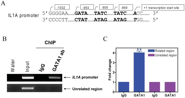
Methodology: Stable clones of CMK cells with decreased GATA1s or Bcl-2 levels were generated by using GATA1- or BCL-2-specific lentivirus shRNAs. In vitro ara-C, daunorubicin, and VP-16 cytotoxicities of the shRNA stable clones were determined by using the Cell Titer-blue reagent. Apoptosis and cell cycle distribution were determined by flow cytometry analysis. Changes in gene transcript levels were determined by gene expression microarray and/or real-time RT-PCR. Changes in protein levels were measured by Western blotting. In vivo binding of GATA1s to IL1A promoter was determined by chromatin immunoprecipitation assays.

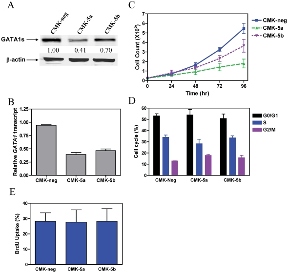
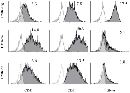
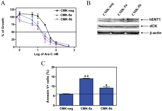
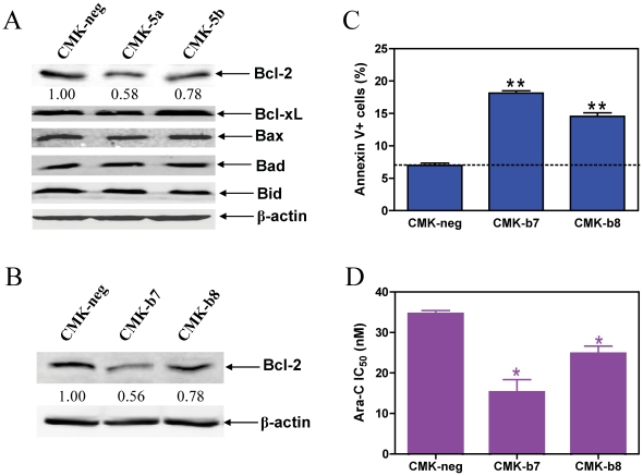
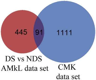
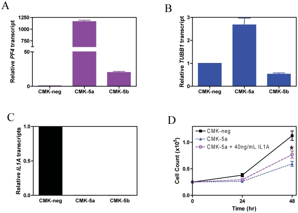
Results: Lentivirus shRNA knockdown of the GATA1 gene in the DS AMkL cell line, CMK (harbors a mutated GATA1 gene and only expresses GATA1s), resulting in lower GATA1s protein levels, promoted cell differentiation towards the megakaryocytic lineage and repressed cell proliferation. Increased basal apoptosis and sensitivities to ara-C, daunorubicin, and VP-16 accompanied by down-regulated Bcl-2 were also detected in the CMK GATA1 shRNA knockdown clones. Essentially the same results were obtained when Bcl-2 was knocked down with lentivirus shRNA in CMK cells. Besides Bcl-2, down-regulation of GATA1s also resulted in altered expression of genes (e.g., IL1A, PF4, and TUBB1) related to cell death, proliferation, and differentiation.

Conclusion: Our results suggest that GATA1s may facilitate leukemogenesis and potentially impact therapeutic responses in DS AMkL by promoting proliferation and survival, and by repressing megakaryocytic lineage differentiation, potentially by regulating expression of Bcl-2 protein and other relevant genes.


Conflict of interest statement

Competing Interests: The authors have declared that no competing interests exist.

Figures

Figure 1
Figure 1. Down-regulation of GATA1s in CMK cells results in impaired cell proliferation.
Expression of GATA1s in two selected subclones, CMK-5a and -5b, in comparison to the negative control (CMK-neg) was verified by Western blotting (panel A) and real-time RT-PCR (panel B). The real-time RT-PCR results were expressed as mean values ± standard errors from 3 independent experiments using the same cDNA preparation and normalized to GAPDH. To establish the doubling times for each shRNA subclone, the CMK-5a, -5b, and –neg sublines were seeded at 2.5×10⁴ cells/ml and counted every 24 h with trypan blue staining (panel C). Cell cycle progression in the CMK-5a, -5b, and –neg sublines was assessed by PI staining and flow cytometry analysis, as described in the Materials and Methods (panel D). DNA content was also assessed in the CMK-5a, -5b, and –neg sublines by incorporating BrdU into DNA and flow cytometry analysis as described in the Materials and Methods (panel E).
Figure 2
Figure 2. Down-regulation of GATA1s in CMK cells results in increased differentiaion toward megakaryocytic lineage.
Megakaryocytic cell surface marker expression in the CMK-5a, -5b, and -neg stable clones was determined by flow cytometry. The unshaded plot represents isotype control. Numbers (isotype ratio) represent the ratio between the quantitative expression value of the cell surface marker and isotype control.
Figure 3
Figure 3. The effects of GATA1s on ara-C sensitivity in CMK cells.
Panel A: The CMK-5a, -5b, and -Neg cells were cultured in complete medium with dialyzed fetal bovine serum in 96-well plates at a density of 8×10⁴ cells/ml, with a range of concentrations of ara-C at 37°C, and viable cell numbers were determined using the Cell Titer-blue reagent and a fluorescent microplate reader. The IC50 values were calculated as the concentrations of drug necessary to inhibit 50% growth compared to control cells cultured in the absence of drugs. The data are presented as mean values ± standard errors from at least 3 independent experiments. Panel B: Soluble proteins from the CMK-5a, -5b, and –neg sublines were subjected to Western blotting and probed by anti-hENT1, -dCK, or β-actin antibodies. Panel C: Basal apoptosis in the CMK-5a, -5b, and –neg cells was determined by annexin V/PI staining and flow cytometry analysis, as described in the Materials and Methods. * and ** indicate p<0.05 and 0.005, respectively.
Figure 4
Figure 4. GATA1s exerts its effects on ara-C sensitivity through modulating Bcl-2 protein levels in CMK cells.
Panel A: Soluble proteins from the CMK-5a, -5b, and –neg sublines were subjected to western blotting and probed by anti-Bcl-2, -Bcl-xL, -Bax, -Bad, -Bid, or β-actin antibodies. Panel B: Expression of Bcl-2 in two selected subclones, CMK-b7 and –b8, in comparison to the negative control (CMK-neg) was verified by Western blotting. Panel C: Basal apoptosis in the CMK-b7, -b8, and –neg cells was determined by annexin V/PI staining and flow cytometry analysis, as described in the Materials and Methods. Panel D: The CMK-b7, -b8, and -Neg cells were cultured in complete medium with dialyzed fetal bovine serum in 96-well plates at a density of 8×10⁴ cells/ml, with a range of concentrations of ara-C at 37°C, and viable cell numbers were determined using the Cell Titer-blue reagent and a fluorescent microplate reader. The IC50 values were calculated as the concentrations of drug necessary to inhibit 50% growth compared to control cells cultured in the absence of drugs. The data are presented as mean values ± standard errors from at least 3 independent experiments. * and ** indicate p<0.05 and 0.005, respectively.
Figure 5
Figure 5. Identification of overlapping genes between the CMK microarray gene set and the previously reported microarray gene set derived from a comparison between DS and non-DS AMkL patient samples.
For each gene set, differentially expressed probes were cross referenced to Entrez Gene IDs. That provided 1111 and 445 Entrex IDs for the CMK gene set and the DS vs non-DS AMkL gene set, respectively. The common set between the 2 groups was found and shown in a Venn representation.
Figure 6
Figure 6. Down-regulation of GATA1s affects PF4, TUBB1, and IL1A expression in CMK cells.
Panels A–C: Transcript levels for PF4 (panel A), TUBB1 (panel B), and IL1A (panel C) in the CMK-5a, -5b, and –neg cells were quantified by real-time RT-PCR as described in the Materials and Methods and results were expressed as mean values ± standard errors from 3 independent experiments using the same cDNA preparation and normalized to GAPDH. Panel D: CMK-5a cells were incubated with or without 40 ng/mL recombinant IL1α protein for up to 48 h and counted in triplicate every 24 h. * indicates p<0.05.
Figure 7
Figure 7. IL1A is a bona fide GATA1s target gene in DS AMkL.
In vivo binding of GATA1s to the putative GATA1 binding sites located in the upstream region of the IL1A gene (panel A) in CMK cells was determined by ChIP assays with use of regular PCR (panel B) and real-time PCR (panel C), as described in Materials and Methods. ** indicates p<0.005.

Similar articles

Cited by

References

    1. Taub JW. Relationship of chromosome 21 and acute leukemia in children with Down syndrome. J Pediatr Hematol Oncol. 2001;23:175–178. - PubMed
    1. Ravindranath Y, Abella E, Krischer JP, Wiley J, Inoue S, et al. Acute myeloid leukemia (AML) in Down's syndrome is highly responsive to chemotherapy: experience on Pediatric Oncology Group AML Study 8498. Blood. 1992;80:2210–2214. - PubMed
    1. Lange BJ, Kobrinsky N, Barnard DR, Arthur DC, Buckley JD, et al. Distinctive demography, biology, and outcome of acute myeloid leukemia and myelodysplastic syndrome in children with Down syndrome: Children's Cancer Group Studies 2861 and 2891. Blood. 1998;91:608–615. - PubMed
    1. Gamis AS, Woods WG, Alonzo TA, Buxton A, Lange B, et al. Increased age at diagnosis has a significantly negative effect on outcome in children with Down syndrome and acute myeloid leukemia: a report from the Children's Cancer Group Study 2891. J Clin Oncol. 2003;21:3415–3422. - PubMed
    1. Athale UH, Razzouk BI, Raimondi SC, Tong X, Behm FG, et al. Biology and outcome of childhood acute megakaryoblastic leukemia: a single institution's experience. Blood. 2001;97:3727–3732. - PubMed

Publication types

MeSH terms