. 2011;6(11):e27542.
doi: 10.1371/journal.pone.0027542. Epub 2011 Nov 14.

EzrA contributes to the regulation of cell size in Staphylococcus aureus

Affiliations

EzrA contributes to the regulation of cell size in Staphylococcus aureus

Ana M Jorge et al. PLoS One. 2011.

Abstract

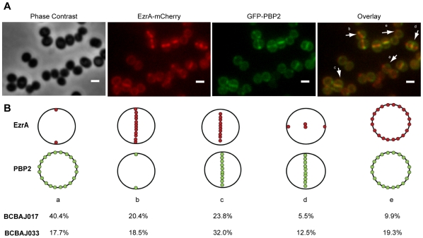
EzrA is a negative regulator of FtsZ in Bacillus subtilis, involved in the coordination between cell growth and cell division and in the control of the cell elongation-division cycle. We have now studied the role of the Staphylococcus aureus homologue of the B. subtilis EzrA protein and shown that it is not essential for cell viability. EzrA conditional and null mutants have an overall increase of the average cell size, compared to wild type strains. In the larger ezrA mutant S. aureus cells, cell division protein FtsZ and the cell wall synthesizing Penicillin Binding Proteins (PBPs) are not properly localized. This suggests that there may be a maximum cell diameter that allows formation of a Z-ring capable of recruiting the other components of the divisome and of driving cytokinesis. We propose that the major role of EzrA in S. aureus is in cell size homeostasis.


Conflict of interest statement

Competing Interests: The authors have declared that no competing interests exist.

Figures

Figure 1
Figure 1. EzrA is not essential in S. aureus.
(A) Bacterial strains with ezrA under the control of inducible Pspac promoter, grown on solid medium (TSA) supplemented or not supplemented with 1 mM IPTG. (a) BCBAJ036 (RN4220 background); (b) BCBAJ031 (NCTC8325-4 background); (c) BCBAJ034 (SH1000 background); (d) BCBAJ035 (Newman background); (e) BCBAJ019 (COL background). (B) Expression of ezrA transcript in control strain BCBHV002 (NCTC8325-4, lacI) and in the inducible strain BCBAJ031 (NCTC8325-4 ezrA:: Pspac -ezrA, lacI), grown in the presence and in the absence of IPTG, quantified by RT-PCR and normalized using 16sRNA. (C) Analysis, by SDS-PAGE, of cell lysates of strains BCBAJ012 (COL ezrA::ezrA-mCherry, lane 1) and BCBAJ024 (COL ezrA::ezrA-mCherry, containing pBCBAJ006 plasmid expressing ezrA antisense RNA) grown in the presence of glucose 0.2% (w/v) to repress antisense RNA transcription (lane 2), or in the presence of xylose 2% (w/v) to induce antisense RNA transcription (lane 3). Bands correspond to fluorescence of EzrA-mCherry detected using a FUJI FLA5100 reader. Integrated density for each band is shown below. (D) Growth curve of wild type strain NCTC8325-4 and null mutant BCBAJ030 (NCTC8325-4 ΔezrA) in liquid medium (TSB).
Figure 2
Figure 2. Absence of EzrA in S. aureus leads to an increase in cell diameter.
Phase contrast images were used to measure the diameter of 600 to 1000 cells of each strain: wild type NCTC8325-4, null mutant BCBAJ030 (NCTC8325-4 ΔezrA), and ezrA conditional mutant BCBAJ031 (NCTC8325-4 ezrA:: Pspac -ezrA, lacI) grown in the presence and in the absence of 1 mM of IPTG inducer.
Figure 3
Figure 3. EzrA-mCherry localizes to the division septa of S. aureus.
Phase contrast (left panels) and fluorescence (right panels) images of BCBAJ025 cells (NCTC8325-4 ezrA::ezrA-mCherry) expressing EzrA-mCherry fusion. Scale bar 1 µm.
Figure 4
Figure 4. FtsZ mislocalizes in large S. aureus cells depleted of EzrA.
(A) Fluorescence images of parental strain BCBHV011 (NCTC8325-4 spa::Pspacftsz-cfp) and ezrA null mutant BCBAJ032 (NCTC8325-4 ΔezrA spa::Pspacftsz-cfp) showing cells expressing FtsZCFP fusion protein. Arrows point to irregular localizations of FtsZ. Scale bar 1 µm. (B) Frequency of cells with septal localization of FtsZ-CFP (class A), fluorescence all over the cytoplasm (class B), double septa (class C) and irregular localizations of FtsZCFP (class D), for BCBHV011 (parental strain; NCTC8325-4 spa::Pspacftsz-cfp) and BCBAJ032 (NCTC8325-4 ΔezrA spa::Pspacftsz-cfp). Cells of null mutant strain BCBAJ032 were divided in two classes - cells with a diameter smaller or larger than 1.75 µm – which were analyzed separately.
Figure 5
Figure 5. Absence of EzrA leads to abnormalities in septum formation.
Electron microscopy images of wild type strain NCTC8325-3 (A), ezrA null mutant strain BCBAJ030 (NCTC8325-4 ΔezrA) (B), ezrA conditional mutant BCBAJ031 (NCTC8325-4 ezrA:: Pspac -ezrA, lacI) grown in the absence of IPTG inducer (C) and in the presence of 1 mM IPTG inducer (D). Arrowheads point to cells in which extra septa started to form. Scale bar 1 µm.
Figure 6
Figure 6. Penicillin Binding Proteins (PBPs) mislocalize in large S. aureus cells depleted of EzrA.
(A) COL (parental strain) and BCBAJ014 (COL ΔezrA) cells labeled with Vancomycin-FL to visualize sites of incorporation of new cell wall. (B) NCTC8325-4 (parental strain) and BCBAJ030 (NCTC8325-4 ΔezrA) cells labeled with Bocillin-FL. Arrows point to cells with irregular localization of PBPs. Scale bar 1 µm. (C) Frequency of cells showing septal localization of PBPs (class A), localization around the membrane (class B) or irregular localization of PBPs (class C) for NCTC8325-4 and BCBAJ030 cells. The later were divided in two classes - cells with a diameter smaller or larger than 1.75 µm – which were analyzed separately.
Figure 7
Figure 7. EzrA interacts with PBPs in a Bacterial Two Hybrid assay.
Plasmid pBCBAJ011 (encoding EzrA-T18) was co-transformed with plasmid p25PBP1, p25PBP2, p25PBP3, p25PBP4, p25PBP2A, pBCBAJ010 (encoding EzrA-T25) and pKT25 (empty vector, negative control) and a 10−2 dilution of each resulting culture was plated. Plasmids p25Zip and p18Zip were used as positive control. Formation of blue colonies indicates putative interaction between cloned proteins, namely between EzrA and itself, PBP1 and PBP2.
Figure 8
Figure 8. EzrA-mCherry and GFP-PBP2 do not colocalize during the entire cell cycle.
(A) From left to right: Phase contrast, EzrA-mCherry fluorescence, GFP-PBP2 fluorescence and overlay of EzrA-mCherry and GFP-PBP2 fluorescence images of strain BCBAJ017 expressing, simultaneously, EzrA-mCherry and GFP-PBP2 in the COL background. Arrows show cells representative for each class (see below). (B) Localization of EzrA-mCherry and GFP-PBP2 was analyzed in 1420 cells for strain BCBAJ017 (COL background) and 802 cells for strain BCBAJ033 (NCTC8325-4 background) which were assigned to the following classes: (a) EzrA-mCherry localized in two spots, corresponding to the beginning of septa formation while GFP-PBP2 is not yet present at mid cell; (b) EzrA-mCherry is present across the entire septum while GFP-PBP2 is present as two dots, corresponding to a ring around the septum; (c) both proteins co-localize at the septum; (d) EzrA-mCherry is starting to localize to the next division site and GFP-PBP2 is still present in the previous septum; (e) both proteins are dispersed at the cell membrane. Red dots represent EzrA-mCherry and green dots GFP-PBP2. Frequency of cells in each class was calculated for BCBAJ017 (COL ezrA::ezrA-mCherry, pbp2::gfp-pbp2) and BCBAJ033 (NCTC8325-4 ezrA::ezrA-mCherry, pbp2::gfp-pbp2).

Similar articles

Cited by

References

    1. Adams DW, Errington J. Bacterial cell division: assembly, maintenance and disassembly of the Z ring. Nat Rev Microbiol. 2009;7:642–653. - PubMed
    1. Bi EF, Lutkenhaus J. FtsZ ring structure associated with division in Escherichia coli. Nature. 1991;354:161–164. - PubMed
    1. Goehring NW, Beckwith J. Diverse paths to midcell: assembly of the bacterial cell division machinery. Curr Biol. 2005;15:R514–526. - PubMed
    1. Weart RB, Levin PA. Growth rate-dependent regulation of medial FtsZ ring formation. J Bacteriol. 2003;185:2826–2834. - PMC - PubMed
    1. Beall B, Lutkenhaus J. Impaired cell division and sporulation of a Bacillus subtilis strain with the ftsA gene deleted. J Bacteriol. 1992;174:2398–2403. - PMC - PubMed

Publication types

MeSH terms