. 2011;6(11):e27560.
doi: 10.1371/journal.pone.0027560. Epub 2011 Nov 14.

SNPs array karyotyping reveals a novel recurrent 20p13 amplification in primary myelofibrosis

Affiliations

SNPs array karyotyping reveals a novel recurrent 20p13 amplification in primary myelofibrosis

Giuseppe Visani et al. PLoS One. 2011.

Abstract

The molecular pathogenesis of primary mielofibrosis (PMF) is still largely unknown. Recently, single-nucleotide polymorphism arrays (SNP-A) allowed for genome-wide profiling of copy-number alterations and acquired uniparental disomy (aUPD) at high-resolution. In this study we analyzed 20 PMF patients using the Genome-Wide Human SNP Array 6.0 in order to identify novel recurrent genomic abnormalities. We observed a complex karyotype in all cases, detecting all the previously reported lesions (del(5q), del(20q), del(13q), +8, aUPD at 9p24 and abnormalities on chromosome 1). In addition, we identified several novel cryptic lesions. In particular, we found a recurrent alteration involving cytoband 20p13 in 55% of patients. We defined a minimal affected region (MAR), an amplification of 9,911 base-pair (bp) overlapping the SIRPB1 gene locus. Noteworthy, by extending the analysis to the adjacent areas, the cytoband was overall affected in 95% of cases. Remarkably, these results were confirmed by real-time PCR and validated in silico in a large independent series of myeloproliferative diseases. Finally, by immunohistochemistry we found that SIRPB1 was over-expressed in the bone marrow of PMF patients carrying 20p13 amplification. In conclusion, we identified a novel highly recurrent genomic lesion in PMF patients, which definitely warrant further functional and clinical characterization.


Conflict of interest statement

Competing Interests: The authors have declared that no competing interests exist.

Figures

Figure 1
Figure 1. Karyoview of training set.
Amplification with LOH (Light blue), Amplification without LOH (Dark blue), Copy Neutral LOH (aUPD, Green), Deletion with LOH (Violet), and Deletion without LOH (Cyclamen) are represented. All the 2,765 lesions recorded in the training set are depicted.
Figure 2
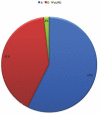
Figure 2. Global distribution of CNVs across PMF genomes.
All the CNVs with relative frequencies are reported: amplifications (both with and without LOH) in blue, acquired uniparental disomy regions (aUPD) in green and deletions (both with and without LOH) in red.
Figure 3
Figure 3. CNVs distribution across chromosomes.
On the x axis all the chromosomes except x, y, and mitochondrion are presented. On the y axis the number of CNVs affecting each chromosome after opportune normalization on chromosome size, is reported. The dotted line represents the median value of CNV per chromosome.
Figure 4
Figure 4. Correspondence between metaphase cytogenetic and SNPs-A karyotyping.
A) Trisomy of chromosome 8 identified in one patient (yellow bar) known to carry the same abnormality basing on metaphase cytogenetic results. Please note two different cases randomly selected and presented as negative controls (arrows). B) Deletion of chromosome 5q identified in one patient (light blue bar) known to carry the same abnormality basing on metaphase cytogenetic results. Please note two different cases randomly selected and presented as negative controls (arrows).
Figure 5
Figure 5. Minimally affected region (MAR) definition.
Genomes from all patients are represented as single rows. MAR (black arrow) was defined as the minimal DNA fragment affected by any imbalance in the majority of cases. Correspondence to SIRPB1 gene was found (red arrow). Please note the occurrence of other abnormalities rather than amplification within the MAR or in the adjacent area in some cases.
Figure 6
Figure 6. Validation of MAR abnormalities TaqMan Copy Number Assay.
Each column represents a single genome (P = patient; C = control). Bars correspond to standard deviations. On the y axis, detected copy number values are presented. C1 = gDNA from Affymetrix; C2 = healthy donor; C3–C7 = patients matched DNA from non neoplastic cells; P1–P10 = patients.
Figure 7
Figure 7. Immunhistochemical expression of SIRPB1.
A–B) In PMF cases not showing 20p13 amplification, SIRPB1 expression is mainly confined to BM macrophages and dendritic cells (B, green arrows) and to scattered myeloid cells (B, red arrows). C–D) By contrast, in cases with 20p13 amplification, SIRPB1 is diffusely expressed by myeloid and megakaryocytic elements with a combined cytoplasmatic and surface expression (D, inset, red arrows). Images are relative to two representative cases out of ten tested (5 per condition). Anti-SIRPB1 immunostaining with the Strept-ABC method and DAB chromogen (brown signal). Original magnifications: a, c ×200; b, d, inset ×400.

References

    1. Thiele J, Kvasnicka HM, Tefferi A, Barosi G, Orazi A, et al. Primary myelofibrosis. In: Swerdlow S, Campo E, Harris NL, Jaffe ES, Pileri SA, et al., editors. WHO Classification of Tumors of the Hematopoietic and Lymphoid Tissue. Lyon: IARC; 2008. pp. 44–47.
    1. Tripodo C, Sangaletti S, Piccaluga P, Prakash S, Franco G, et al. The bone marrow stroma in hematological neoplasms—a guilty bystander. Nature Reviews Clinical Onclogy. 2011 in press. - PMC - PubMed
    1. Tripodo C, Di Bernardo A, Ternullo MP, Guarnotta C, Porcasi R, et al. CD146(+) bone marrow osteoprogenitors increase in the advanced stages of primary myelofibrosis. Haematologica. 2009;94:127–130. - PMC - PubMed
    1. Stein BL, Moliterno AR. Primary myelofibrosis and the myeloproliferative neoplasms: the role of individual variation. Jama. 2010;303:2513–2518. - PubMed
    1. Tefferi A. Novel mutations and their functional and clinical relevance in myeloproliferative neoplasms: JAK2, MPL, TET2, ASXL1, CBL, IDH and IKZF1. Leukemia. 2010;24:1128–1138. - PMC - PubMed

Publication types

MeSH terms