. 2011;6(11):e27586.
doi: 10.1371/journal.pone.0027586. Epub 2011 Nov 16.

Activation of GPR4 by acidosis increases endothelial cell adhesion through the cAMP/Epac pathway

Affiliations

Activation of GPR4 by acidosis increases endothelial cell adhesion through the cAMP/Epac pathway

Aishe Chen et al. PLoS One. 2011.

Abstract

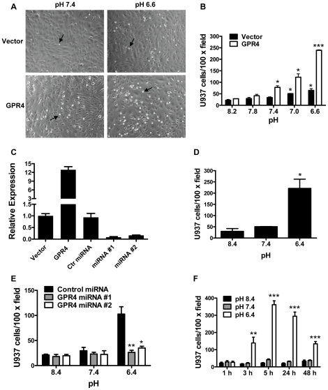
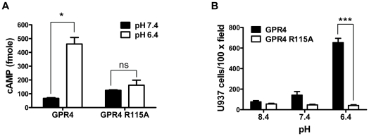
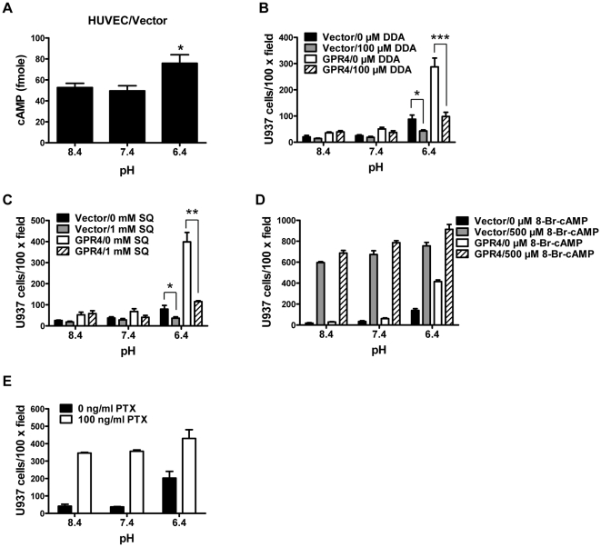
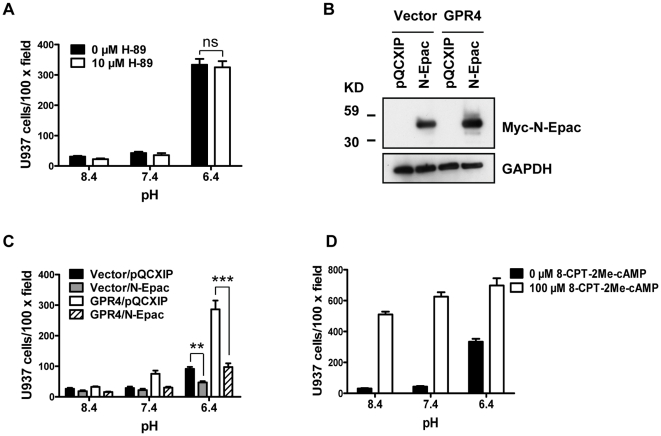
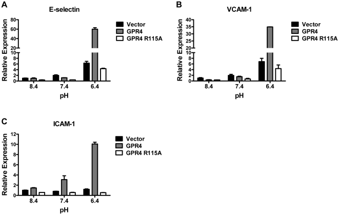
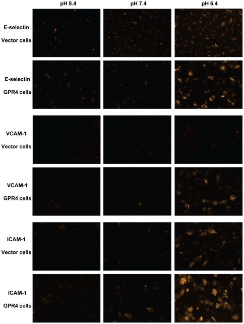
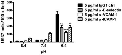
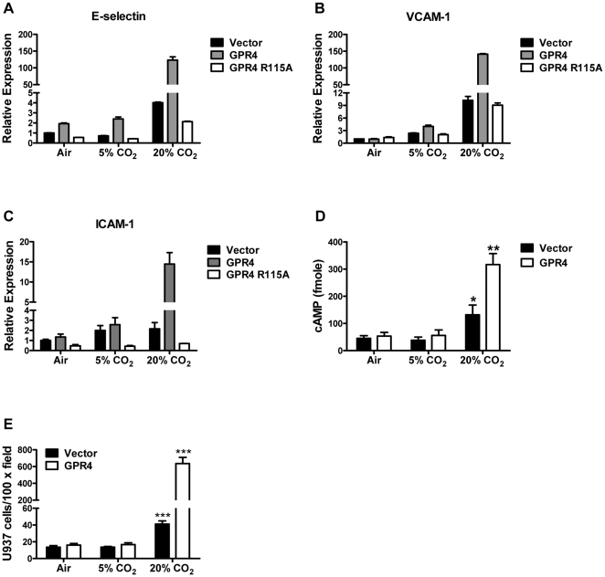
Endothelium-leukocyte interaction is critical for inflammatory responses. Whereas the tissue microenvironments are often acidic at inflammatory sites, the mechanisms by which cells respond to acidosis are not well understood. Using molecular, cellular and biochemical approaches, we demonstrate that activation of GPR4, a proton-sensing G protein-coupled receptor, by isocapnic acidosis increases the adhesiveness of human umbilical vein endothelial cells (HUVECs) that express GPR4 endogenously. Acidosis in combination with GPR4 overexpression further augments HUVEC adhesion with U937 monocytes. In contrast, overexpression of a G protein signaling-defective DRY motif mutant (R115A) of GPR4 does not elicit any increase of HUVEC adhesion, indicating the requirement of G protein signaling. Downregulation of GPR4 expression by RNA interference reduces the acidosis-induced HUVEC adhesion. To delineate downstream pathways, we show that inhibition of adenylate cyclase by inhibitors, 2',5'-dideoxyadenosine (DDA) or SQ 22536, attenuates acidosis/GPR4-induced HUVEC adhesion. Consistently, treatment with a cAMP analog or a G(i) signaling inhibitor increases HUVEC adhesiveness, suggesting a role of the G(s)/cAMP signaling in this process. We further show that the cAMP downstream effector Epac is important for acidosis/GPR4-induced cell adhesion. Moreover, activation of GPR4 by acidosis increases the expression of vascular adhesion molecules E-selectin, VCAM-1 and ICAM-1, which are functionally involved in acidosis/GPR4-mediated HUVEC adhesion. Similarly, hypercapnic acidosis can also activate GPR4 to stimulate HUVEC adhesion molecule expression and adhesiveness. These results suggest that acidosis/GPR4 signaling regulates endothelial cell adhesion mainly through the G(s)/cAMP/Epac pathway and may play a role in the inflammatory response of vascular endothelial cells.


Conflict of interest statement

Competing Interests: Li V. Yang is the applicant for a patent which includes the utilization of GPR4 in the treatment of inflammation, related to the data described in this manuscript. This does not alter the authors' adherence to all the PLoS ONE policies on sharing data and materials.

Figures

Figure 1
Figure 1. Acidosis/GPR4-induced adhesion of HUVEC cells.
(A) HUVECs stably expressing the MSCV-huGPR4-IRES-GFP construct (GPR4) or the MSCV-IRES-GFP control vector (Vector) were grown as a flat monolayer and treated with EGM-2/HEM media at varying pH for either 5 h or 15 h (overnight) which showed similar results. Then, 6×10⁴ cells/well of U937 monocytic cells were added to adhere with HUVECs for 1 h at pH 7.4, and the static cell adhesion assay was performed as described in the “Materials and Methods”. After the adhesion assay, the attached U937 cells were readily detected as small, round and reflectile cells under an inverted microscope with a 10× objective. Arrows indicate attached U937 cells. (B) HUVECs stably overexpressing GPR4 or the control vector were treated with different pHs as indicated. The cell adhesion assay was then performed. *, P<0.05; ***, P<0.001; compared with the pH 8.2 group. (C) HUVECs were transduced with various genetic constructs as indicated. Total RNA was isolated and mRNA levels of GPR4 were determined by real-time RT-PCR. Values were normalized to the housekeeping gene GAPDH. The expression level of GPR4 in HUVECs with the control vector was set as 1. (D) HUVECs stably overexpressing the mouse GPR4-GFP fusion gene were treated with different pHs as indicated, and then the cell adhesion assay was performed. Error bars are the mean ± SEM. *, P<0.05, compared with pH 8.4. (E) HUVECs were transduced with two GPR4 miRNAs or control miRNA, followed by pH treatment, and then the cell adhesion assay was performed. *, P<0.05; **, P<0.01; compared with the control miRNA group at pH 6.4. (F) HUVEC/GPR4 cells were treated with different pHs for various lengths of time as indicated, and then the cell adhesion assay was performed. **, P<0.01; ***, P<0.001; compared with the 1 h group at pH 6.4. The results are depicted as the mean ± SEM and representative of more than two independent experiments.
Figure 2
Figure 2. G protein signaling is indispensible for acidosis/GPR4-induced HUVEC adhesion.
(A) HUVECs were transduced with GPR4 or the GPR4 R115A mutant expression vector. The intracellular cAMP level in HUVECs was determined by the cAMP assay as described under “Materials and Methods”. The total intracellular cAMP was calculated according to the cAMP standard measured in parallel. *, P<0.05; ns, not significant (P>0.05). (B) HUVEC cells as described in (A) were treated with indicated pHs and the cell adhesion assay was performed as described under “Materials and Methods”. ***, P<0.001. The results are representative of two independent experiments. Error bars are the mean ± SEM.
Figure 3
Figure 3. Involvement of cAMP in acidosis/GPR4-induced HUVEC adhesion.
(A) The intracellular cAMP level in pH-treated HUVEC/Vector cells was determined by the cAMP assay as described in the “Materials and Methods”. *, P<0.05; compared with pH 8.4. (B) HUVECs stably overexpressing GPR4 or the control vector were pretreated with vehicle or 100 µM 2′,5′-dideoxyadenosine (DDA) for 1 h, followed by the treatment with indicated pH media containing vehicle or 100 µM DDA for 5 h, and the cell adhesion assay was performed as described in the “Materials and Methods”. *, P<0.05; ***, P<0.001. (C) HUVECs stably overexpressing GPR4 or the control vector were pretreated with vehicle or SQ 22536 (1 mM) for 1 h, followed by the treatment with indicated pH media containing vehicle or 1 mM SQ 22536 for 5 h, and then the cell adhesion assay was performed. *, P<0.05; **, P<0.01. (D) HUVECs stably overexpressing GPR4 or the control vector were treated with indicated pH media containing vehicle or 500 µM 8-bromo-cAMP for 15 h (overnight), and then the cell adhesion assay was performed. (E) HUVEC/GPR4 cells were treated with indicated pH media containing vehicle or 100 ng/ml PTX for 15 h, and then the cell adhesion assay was performed. All the above results are representative of at least two independent experiments. Error bars are the mean ± SEM.
Figure 4
Figure 4. Epac is important for HUVEC adhesion induced by acidosis/GPR4.
(A) HUVEC/GPR4 cells were pretreated with vehicle or H-89 (10 µM) for 1 h. Cells were then treated with indicated pH media containing vehicle or 10 µM H-89 for 5 to 15 h (overnight), and the cell adhesion assay was performed as described under “Materials and Methods”. ns, not significant (P>0.05). (B) HUVEC/Vector or HUVEC/GPR4 cells were stably transduced with the myc-tagged Epac dominant-negative construct N-Epac or the control vector pQCXIP. Total proteins were extracted from HUVEC cells and subject to Western blot analysis for myc-N-Epac with anti-myc antibodies. GAPDH was used as a loading control. (C) HUVEC/Vector or HUVEC/GPR4 cells were stably transduced with the dominant-negative N-Epac or the control vector pQCXIP. Cells were treated with indicated pH media for 5 or 15 h and then the cell adhesion assay was performed. **, P<0.01; ***, P<0.001. (D) HUVEC/GPR4 cells were treated with indicated pH media containing vehicle or 100 µM 8-CPT-2Me-cAMP for 15 h, and then the cell adhesion assay was performed. All the above results are representative of two or more independent experiments. Error bars are the mean ± SEM.
Figure 5
Figure 5. Real-time RT-PCR of adhesion molecules E-selectin, VCAM-1, and ICAM-1.
HUVECs transduced with the control vector, GPR4 or GPR4 R115A mutant were treated with EGM-2/HEM media at pH 8.4, 7.4, or 6.4 for 5 h. Total RNAs were isolated and reverse transcribed. Real-time RT-PCR quantification of mRNA levels of E-selectin (A), VCAM-1 (B), and ICAM-1 (C) was performed in duplicate. Values were normalized to the housekeeping gene GAPDH. The expression level of the target gene in HUVEC/Vector cells at pH 8.4 was set as 1. The results are representative of at least two independent experiments. Error bars are the mean ± SEM.
Figure 6
Figure 6. Immunofluorescence staining of E-selectin, VCAM-1 and ICAM-1 in HUVEC cells.
After treated with EGM-2/HEM media at pH 8.4, 7.4, or 6.4 for 5 h, HUVEC/Vector and HUVEC/GPR4 cells were fixed with 4% paraformaldehyde, incubated with E-selectin, VCAM-1 or ICAM-1 primary antibody, Rhodamine Red-conjugated secondary antibody, and then detected under a fluorescence microscope (10× objective). For an accurate comparison of fluorescence signals, each group of images (e.g. E-selectin/Vectors cells, pH 8.4, pH 7.4, and pH 6.4) was taken with the same exposure time. The results are representative of three independent experiments.
Figure 7
Figure 7. Blockade of E-selectin, VCAM-1 or ICAM-1 reduces the adhesion of HUVECs with U937 monocytic cells.
HUVECs stably overexpressing GPR4 were treated with different pHs as indicated for 5 to 15 h, followed by 30 min incubation with pH 7.4 medium containing 5 µg/ml IgG1 control antibody, anti-E-selectin, anti-VCAM-1, or anti-ICAM-1 antibody. The cell adhesion assay was then performed as described under “Materials and Methods”. **, P<0.01; ***, P<0.001; compared with the IgG1 control group at pH 6.4. The results are representative of two independent experiments. Error bars are the mean ± SEM.
Figure 8
Figure 8. Activation of GPR4 by hypercapnic acidosis stimulates HUVEC adhesion molecule expression, cAMP production, and adhesiveness.
(A–C) HUVEC/Vector, HUVEC/GPR4, or HUVEC/GPR4 R115A cells were treated with ambient air, 5% CO2, or 20% CO2-buffered EGM-2 pH media for 5 h. Total RNA was isolated and mRNA levels of E-selectin (A), VCAM-1 (B), and ICAM-1 (C) were determined by real-time RT-PCR. Values were normalized to the housekeeping gene GAPDH. The expression level of the target gene in HUVEC/Vector cells under ambient air (∼pH 8.4) was set as 1. (D) The intracellular cAMP level in HUVEC/Vector and HUVEC/GPR4 cells treated with the CO2-buffered pH media was determined by the cAMP assay as described in the “Materials and Methods”. *, P<0.05; **, P<0.01; compared with the 5% CO2 group. (E) HUVEC/Vector and HUVEC/GPR4 cells were treated with the CO2-buffered pH media as indicated for 5 h. The cell adhesion assay was performed as described in the “Materials and Methods”. ***, P<0.001; compared with the 5% CO2 group. All the above results are representative of at least two independent experiments. Error bars are the mean ± SEM.

Similar articles

Cited by

References

    1. Gatenby RA, Gillies RJ. Why do cancers have high aerobic glycolysis? Nat Rev Cancer. 2004;4:891–899. - PubMed
    1. Hunt JF, Fang K, Malik R, Snyder A, Malhotra N, et al. Endogenous airway acidification. Implications for asthma pathophysiology. Am J Respir Crit Care Med. 2000;161:694–699. - PubMed
    1. Koul PB. Diabetic ketoacidosis: a current appraisal of pathophysiology and management. Clin Pediatr (Phila) 2009;48:135–144. - PubMed
    1. Lardner A. The effects of extracellular pH on immune function. J Leukoc Biol. 2001;69:522–530. - PubMed
    1. Nedergaard M, Kraig RP, Tanabe J, Pulsinelli WA. Dynamics of interstitial and intracellular pH in evolving brain infarct. Am J Physiol. 1991;260:R581–588. - PMC - PubMed

Publication types

MeSH terms