. 2011;6(11):e27696.
doi: 10.1371/journal.pone.0027696. Epub 2011 Nov 15.

The identification of a small molecule compound that reduces HIV-1 Nef-mediated viral infectivity enhancement

Affiliations

The identification of a small molecule compound that reduces HIV-1 Nef-mediated viral infectivity enhancement

Nopporn Chutiwitoonchai et al. PLoS One. 2011.

Abstract

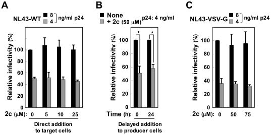
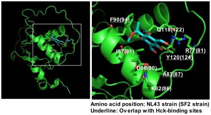
Nef is a multifunctional HIV-1 protein that accelerates progression to AIDS, and enhances the infectivity of progeny viruses through a mechanism that is not yet understood. Here, we show that the small molecule compound 2c reduces Nef-mediated viral infectivity enhancement. When added to viral producer cells, 2c did not affect the efficiency of viral production itself. However, the infectivity of the viruses produced in the presence of 2c was significantly lower than that of control viruses. Importantly, an inhibitory effect was observed with Nef(+) wild-type viruses, but not with viruses produced in the absence of Nef or in the presence of proline-rich PxxP motif-disrupted Nef, both of which displayed significantly reduced intrinsic infectivity. Meanwhile, the overexpression of the SH3 domain of the tyrosine kinase Hck, which binds to a PxxP motif in Nef, also reduced viral infectivity. Importantly, 2c inhibited Hck SH3-Nef binding, which was more marked when Nef was pre-incubated with 2c prior to its incubation with Hck, indicating that both Hck SH3 and 2c directly bind to Nef and that their binding sites overlap. These results imply that both 2c and the Hck SH3 domain inhibit the interaction of Nef with an unidentified host protein and thereby reduce Nef-mediated infectivity enhancement. The first inhibitory compound 2c is therefore a valuable chemical probe for revealing the underlying molecular mechanism by which Nef enhances the infectivity of HIV-1.


Conflict of interest statement

Competing Interests: The authors have declared that no competing interests exist.

Figures

Figure 1
Figure 1. The effect of 2c on the infectivity of NL43 wild-type and Nef-defective mutant viruses.
(A) The chemical structure of 2c. (B) The infectivity of the NL43 wild-type (WT) and Nef-defective mutant (ΔNef) viruses to the target TZM-bl cells was compared by varying the concentration of p24 Gag protein and is expressed as a percentage of the value for the sample on the far left. Data are shown as the mean±SD of triplicate assays and are representative of two independent experiments with similar results. (C) 2c was added to 293 cells producing NL43-WT or ΔNef viruses at the indicated concentrations for 2 days, and the concentration of p24 Gag protein in the cell supernatants was determined by ELISA (bar graph). Data are shown as the mean±SD of triplicate assays and are representative of two independent experiments with similar results. Alternatively, the producer cells were lysed and analyzed for the expression of Gag and Nef by Western blotting (lower blots). The actin blot was used as a loading control. (D) The infectivity of NL43-WT (upper) or ΔNef viruses (lower) produced by 293 cells in the absence or presence of the indicated concentrations of 2c was determined using TZM-bl cells as the target cells. The WT and ΔNef viruses were inoculated by changing the p24 concentration (2 or 4 ng/ml and 8 or 16 ng/ml for the WT and ΔNef viruses, respectively) so that the two viruses were similarly infective to the target cells. Infectivity is expressed as a percentage of the value for the sample on the far left. Data are shown as the mean±SD of triplicate assays and are representative of three independent experiments with similar results. *p<0.05.
Figure 2
Figure 2. Several features of the activity of 2c on viral infectivity.
(A) 2c was added to the target TZM-bl cells at the indicated concentrations together with the NL43 wild-type (WT) viruses produced in the absence of 2c. The amount of p24 inoculated was 4 or 8 ng/ml. The infectivity is expressed as a percentage of the value for the sample on the far left. (B) 2c (50 µM) or the control DMSO was added to the producer 293 cells immediately after transfection (0 h) or 24 h after transfection of the NL43 WT plasmid. The infectivity of the viruses was determined using TZM-bl cells and is expressed as a percentage of the value for the sample on the far left. The amount of p24 inoculated was 4 ng/ml. (C) 2c (50 or 75 µM) or the control DMSO was added to the producer 293 cells immediately after co-transfection of Env-defective NL43 plasmid and VSV-G expression plasmid. The infectivity of the pseudotyped viruses was determined using TZM-bl cells and is expressed as a percentage of the value for the sample on the far left. The amount of p24 inoculated was 4 or 8 ng/ml. (A–C) Data are shown as the mean±SD of triplicate assays and are representative of two independent experiments with similar results. *p<0.05.
Figure 3
Figure 3. The effect of 2c on the replication of HIV-1.
(A) Jurkat cells were infected with either the NL43 wild-type (WT) or Nef-defective (ΔNef) viruses at the indicated concentrations of p24, and cultured in the presence (50 µM) or absence of 2c. AZT was also used at 5 µM. The concentration of p24 in the supernatants (at day 5, 7 or 9) was determined by ELISA. Data are shown as the mean±SD of triplicate assays and are representative of two independent experiments with similar results. (B) Peripheral blood monocyte-derived macrophages were obtained from two different donors, infected with either the JRFL wild-type (WT) or Nef-defective (ΔNef) viruses at the indicated concentrations of p24, and cultured in the presence (50 µM) or absence of 2c. AZT was also used at 5 µM. The concentration of p24 in the supernatants (at day 6, 9 or 12) was determined by ELISA. Data are shown as the mean±SD of triplicate assays. *p<0.05.
Figure 4
Figure 4. The effect of 2c on the infectivity of NL43 viruses with point amino acid mutations in Nef.
(A) The infectivity of the indicated NL43 viruses produced by 293 cells in the absence or presence of 50 µM 2c was determined using TZM-bl cells as the target cells and is expressed as a percentage of the value for the sample on the far left. The amount of p24 inoculated was 10 ng/ml. Wild-type (WT), Nef-defective (ΔNef), or viruses with the indicated amino acid point mutations in Nef (R77A, K82A, D86A, F90A, or G119L) were used. (B) The infectivity of NL43 viruses with the G119L mutation in Nef or ΔNef viruses produced by 293 cells in the absence or presence of 50 µM 2c was determined using TZM-bl cells as the target cells and is expressed as a percentage of the value for the sample on the far left. The amount of p24 inoculated was 2, 8, or 32 ng/ml. (A, B) Data are shown as the mean±SD of triplicate assays and are representative of two independent experiments with similar results. *p<0.05.
Figure 5
Figure 5. The effect of 2c on the infectivity of SF2 wild-type, Nef-defective, and Nef PxxP motif-disrupted viruses.
(A) The infectivity of the SF2 wild-type (WT), Nef-defective (ΔNef), and Nef PxxP motif-disrupted viruses (AxxA) was compared by inoculating them into the target TZM-bl cells at a concentration of 8 ng/ml p24 and is expressed as a percentage of the value for the sample on the far left. Data are shown as the mean±SD of triplicate assays and are representative of two independent experiments with similar results. *p<0.05. (B) 2c was added to 293 cells producing SF2-WT, ΔNef, or AxxA viruses at the indicated concentrations for 2 days, and the concentration of p24 Gag protein in the supernatants was determined by ELISA (bar graph). Data are shown as the mean±SD of triplicate assays and are representative of two independent experiments with similar results. Alternatively, the producer cells were lysed and analyzed for the expression of Gag, Nef, and Vif by Western blotting (lower blots). The actin blot was used as a loading control. (C) The infectivity of SF2-WT (top), AxxA (middle), or ΔNef viruses (bottom) produced by 293 cells in the absence or presence of the indicated concentrations of 2c was determined using TZM-bl cells as the target cells. The WT, AxxA, and ΔNef viruses were inoculated by changing the concentration of p24 (8 ng/ml, 16 ng/ml, and 32 ng/ml for WT, AxxA and ΔNef viruses, respectively) so that these viruses were similarly infective to the target cells. Infectivity is expressed as a percentage of the value for the sample on the far left. In the top panel, the infectivity values of the AxxA and ΔNef viruses produced at the same concentration of p24 (i.e., 8 ng/ml) are also shown as a reference. In the middle panel, the infectivity values of the ΔNef viruses produced at the same concentration of p24 (i.e., 16 ng/ml) are also shown. Data are shown as the mean±SD of triplicate assays and are representative of three independent experiments with similar results. *p<0.05.
Figure 6
Figure 6. The effects of the overexpression of mutant forms of Hck on viral infectivity.
(A) The mutant forms of Hck used are shown schematically. HckN lacks the kinase domain and the two intra-molecular interactions present in the wild-type (WT) Hck. HckN-based HckN-R151S and HckN-W93F have amino acid substitutions in their SH2 and SH3 domain, respectively. (B) The 293 cells were transfected with the NL43 wild-type proviral plasmid or co-transected with the indicated amount of plasmid (HckN, HckN-R151S, or HckN-W93F). The infectivity of the viruses produced in the supernatants was determined using TZM-bl cells as the target cells and is expressed as a percentage of the value for the sample on the far left (bar graph). The amount of p24 inoculated was 8 ng/ml. Alternatively, the producer 293 cells were lysed and analyzed for the expression of the mutant Hck proteins by Western blotting (blot).
Figure 7
Figure 7. The effect of 2c on binding between Nef and Hck.
(A) The Nef proteins fused to GST are shown schematically. In addition to the wild-type (WT) SF2 and NL43 strain Nef, the NL43-TR mutant, which contained a T71R amino acid substitution, and another NL43 AxxA mutant, in which the PxxP motif was disrupted (P72A and P75A substitutions), were used. (B) The resins to which the control GST or indicated GST-Nef fusion proteins were bound were incubated with the lysates of 293 cells expressing the indicated Hck protein. The amount of Hck bound to the resins was determined by Western blotting (pull-down assay). To confirm the equal expression of these Hck proteins in the 293 cells, equal amounts of each cell lysate were analyzed (Input Hck). Moreover, the amounts of the GST and GST-Nef fusion proteins bound to the resins were verified by the elution from the resins followed by SDS-PAGE/Coomassie brilliant blue (CBB) staining. (C) Three different competitive pull-down assays were performed. In the experiment shown in the left panel, the resins to which the GST-SF2 Nef fusion proteins were bound were incubated with the lysates obtained from the 293 cells expressing the wild-type Hck for 3 h, and then 2c was added to the mixture at the indicated concentration. In the experiment shown in the middle panel, the resins to which the GST-SF2 Nef fusion proteins were bound were incubated with the lysates of 293 cells expressing the wild-type Hck and the indicated concentration of 2c. In the experiment shown in the right panel, the resins to which the GST-SF2 Nef fusion proteins were bound were first incubated with the indicated concentration of 2c for 4 h and then washed to remove unbound 2c. Then, the resins were incubated with the lysates of 293 cells expressing the wild-type Hck. The amount of Hck bound to the resins was determined by Western blotting (upper blots). The GST-Nef blot was used as a loading control (lower blots). Data shown are representative of two independent experiments with similar results.
Figure 8
Figure 8. The effect of 2c on the binding between Nef PxxP motif-containing peptides and Hck.
(A) The Nef peptide fused to GST is shown schematically. The 20 amino acid peptide derived from the PxxP motif of SF2 Nef was used (the proline residues are underlined). The resins to which the control GST, GST-SF2 Nef-PxxP peptides (SF2-PxxP), or GST-intact SF2 Nef (SF2-WT) fusion proteins were bound were incubated with the lysates of 293 cells expressing the indicated Hck protein. The amount of Hck bound to each resin was determined by Western blotting (Pull-down). To verify the equal expression of these Hck proteins in the 293 cells, equal amounts of each cell lysate were analyzed (Input Hck). Moreover, the amounts of the GST and GST-Nef fusion proteins bound to the resins were verified by eluting from the resins followed by SDS-PAGE/Coomassie brilliant blue (CBB) staining. (B) Two different competitive pull-down assays were performed. In the experiment shown in the left panel, the resins to which the GST-SF2 Nef-PxxP peptides were bound were incubated with the lysates of 293 cells expressing the wild-type Hck and the indicated concentration of 2c. In the experiment shown in the right panel, the resins to which the GST-SF2 Nef-PxxP peptides were bound were incubated with the indicated concentrations of 2c for 4 h and then washed to remove unbound 2c. Then, the resins were incubated with the lysates of 293 cells expressing the wild-type Hck. The amount of Hck bound to the resins was determined by Western blotting (upper blots). The GST-Nef blot was used as a loading control (lower blots). Data shown are representative of two independent experiments with similar results.
Figure 9
Figure 9. The 2c-Nef docking model.
The amino acids that are predicted to be involved in the interaction between Nef and 2c are indicated. The positions of these amino acids in the NL43 strain and SF2 strain are shown. The amino acids predicted to interact with the Hck SH3 domain are underlined .
Figure 10
Figure 10. A model of the inhibitory effect of 2c.
Both 2c and the Hck SH3 domain bind directly to Nef and reduce viral infectivity, probably by inhibiting the interaction of Nef with an unidentified cellular protein(s).

Similar articles

Cited by

References

    1. Fackler OT, Baur AS. Live and let die: Nef functions beyond HIV replication. Immunity. 2002;16:493–497. - PubMed
    1. Peterlin BM, Trono D. Hide, shield and strike back: how HIV-infected cells avoid immune eradication. Nat Rev Immunol. 2003;3:97–107. - PubMed
    1. Foster JL, Garcia JV. HIV-1 Nef: at the crossroads. Retrovirology. 2008;5:84. - PMC - PubMed
    1. Malim MH, Emerman M. HIV-1 accessory proteins–ensuring viral survival in a hostile environment. Cell Host Microbe. 2008;3:388–398. - PubMed
    1. Deacon NJ, Tsykin A, Solomon A, Smith K, Ludford-Menting M, et al. Genomic structure of an attenuated quasi species of HIV-1 from a blood transfusion donor and recipients. Science. 1995;270:988–991. - PubMed

Publication types

MeSH terms

Substances