. 2008 Nov;1(1):1-14.
doi: 10.1055/s-0028-1098968.

Chin ptosis: classification, anatomy, and correction

Affiliations

Chin ptosis: classification, anatomy, and correction

Evan S Garfein et al. Craniomaxillofac Trauma Reconstr. 2008 Nov.

Abstract

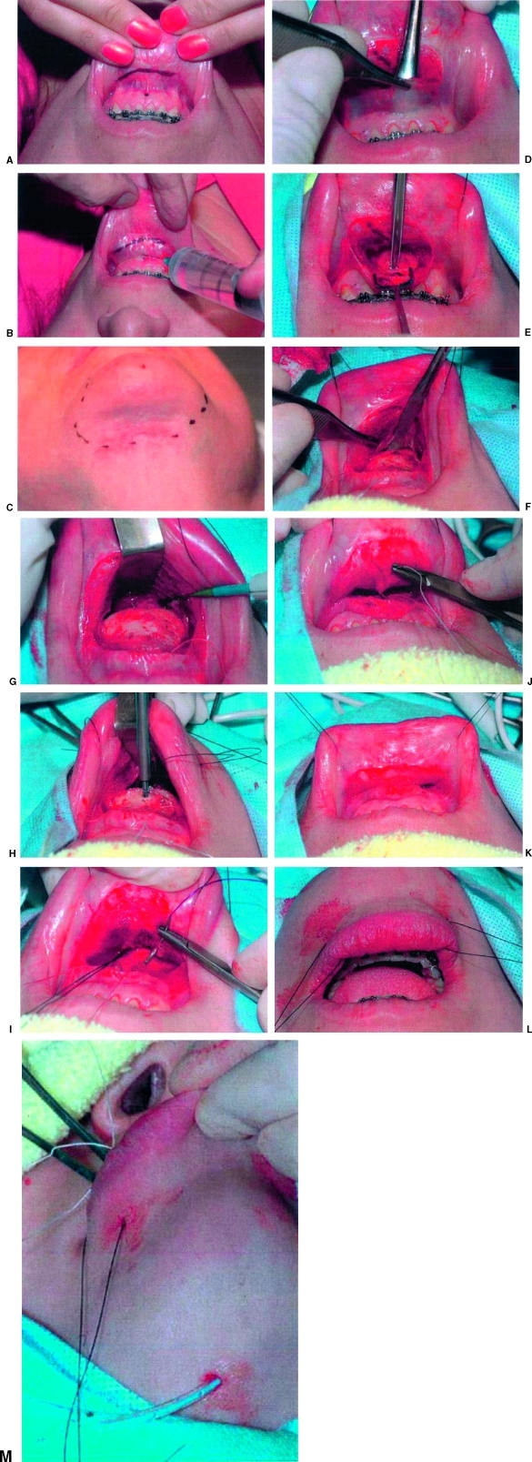
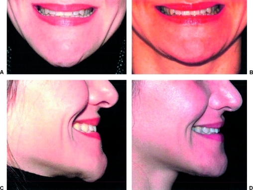
For years, the notion of chin ptosis was somehow integrated with the concept of witch's chin. That was a mistake on many levels because chin droop has four major causes, all different and with some overlap. With this article, the surgeon can quickly diagnose which type and which therapeutic modality would work best. In some cases the problem is a simple fix, in others the droop can only be stabilized, and in the final two, definite corrective procedures are available. Of note, in certain situations two types of chin ptosis may overlap because both the patient and the surgeon may each contribute to the problems. For example, in dynamic ptosis, a droop that occurs with smile in the unoperated patient can be exacerbated and further produced by certain surgical methods also. This paper classifies the variations of the problems and explains the anatomy with the final emphasis on long-term surgical correction, well described herein. This article is the ninth on this subject and a review of them all would be helpful (greatly) for understanding the enigmas of the lower face.

Keywords: Lip incompetence; chin droop; chin ptosis; mentalis muscle; witch's chin.


Figures

Figure 1
Figure 1
Patient with Bell's palsy. (A) Note atrophy of mentalis muscle with loss of bulk on the patient's right. (B) Note inability to elevate lower lip on affected side (right side) and loss of dimpling with attempted contraction of mentalis muscles.
Figure 2
Figure 2
(A) Arrow points to the most medial of the lip depressors, which consist of triangularis, quadratus, anterior platysma. Shiny tissues medially are the mentalis insertions into the chin pad. (B) The level at which the orbicularis oris and the mentalis muscles intersect leads, in this cadaver, to a high labiomental fold (arrowhead). The distinctiveness of the fold is determined by the amount of overlap. The upper part of the mentalis muscle contains horizontally directed fibers that stabilize the lower lip (arrow). The oblique lower fibers allow the lip to pout.
Figure 3
Figure 3
Illusory ptosis: (A) In this unoperated patient, a crease exists that extends on each side laterally onto the face. Even with smiling, the pad and crease remain stable. (B) Postoperative image following creation of a de-epithelialized flap placed under the excess crease, placement of a small implant, and lateral undermining. The contour is improved.
Figure 4
Figure 4
The balling phenomenon is caused by the contraction of the scar under the mentalis muscles. (A,B) When an implant is removed and nothing is done, the contraction may provide some residual projection, but lip compression will show the dimpling or reduction of the pad size.
Figure 5
Figure 5
(A) Vestibuloplasty with replacement by a skin graft made central lip closure impossible and drooling occurred constantly. (B) Note lower central lip at rest.
Figure 6
Figure 6
(A,B) Unoperated dynamic ptosis. These patients always have a horizontal upper lip with smile. The zygomatic muscles fail to lift the commissures, allowing risorius dominance with smile, effacing the soft tissue against the bone and causing the chin pad to move inferiorly.
Figure 7
Figure 7
(A) Developmental ptosis due to denture trauma on the alveolar bone. (B) The upper mentalis fibers originate from the top of the ridge. (C) During surgery (another patient), the fibers can be seen coming directly from the top of the ridge (arrows). (D) In a cadaver, the side view shows the mentalis origin coming from the top of the edentulous ridge. Note: If a woodwind musician loses the ability to purse his or her lips, this results in loss of embouchure and playing is made difficult.
Figure 8
Figure 8
Although some patients will show incisors at rest (A and C), all have a low, scarred sulcus (B and D). Patients complain that they must think to close their lips, straining the lower mentalis, and causing dimpling or fasciculations at the chin pad.
Figure 9
Figure 9
(A) Incision is made on the lip. The dot shows the height of the mentalis reinsertion. (B) The area between the incision and the dot is tumesced for ease of proximal flap elevation. (C) The area of submental undermining is marked and injected with a small amount of extra anesthetic placed on one side for drain placement. (D) After the incision extends from “nerve to nerve,” the proximal tumesced flap is elevated to the lower attached gingiva. If the patient has periodontal disease, the reinsertion point may need to be lower. (E) An absorbable anchor is placed between the central incisors (the anchor can be placed later). (F) Leaving the lower, avascular origin of the mentalis attached to the bone below the anchor, the symphysis is exposed from bicuspid to bicuspid. The menton area periosteum is released from side to side to approach the submental region. (G) Submental, wide, supraplatysmal undermining will allow the neck, as well as the chin pad, to move up. (H) A lower, permanent, larger anchor is placed to suture to the lower mentalis fibers. If hardware or a porous implant is present, the suture may go from the muscle to the prosthesis. A drain is placed in the submental area. (I) The dark suture is the lower anchor suture. Now, semipermanent sutures are placed from the muscle to the drifted mentalis remnant. (J) The final suture comes from the upper anchor. This double-armed suture is placed as a figure 8, while elevating the chin from below. (K) Closure of the mucosa is performed with a central 4-0 chromic suture and then a water-tight, running suture. (L) The lip now rests easily above the level of the lower incisors. A dressing is placed over the drain and jaw put so the upper part rests below the lip. (M) A view of the dependent drain exiting the submental area is seen. The drain is removed in 1 or 2 days.
Figure 10
Figure 10
Case 1. This patient presented with a high labiomental fold and removal of a large chin implant. The combination of a high, indistinct fold and high chin pad percentage made her chin appear enormous after implant placement. After implant removal, the capsule contracted and the overlying stretched muscle and skin drooped. This led to iatrogenic dynamic ptosis and “balling.” Repair involved defatting the excess lower tissue and reorientation of the lower mentalis to the platysma in a “vest over pants” fashion. (A and C) Preoperative photos. (B and D) Postoperative photos.
Figure 11
Figure 11
Case 2. This woman presented with dynamic ptosis and a deficient chin sagittally. No excision of skin was done until the implant was placed and the area defatted. The final result shows the shorter chin with smile.
Figure 12
Figure 12
Case 3. This woman presented with dynamic ptosis and a previous chin implant that was placed too high. This accentuated the labiomental fold, was uncomfortable, and was visible below the fold with smile. The neck was also untreated with excess fat and bands. Surgery was performed via a submental approach; the neck was defatted and a Feldman corset platysmaplasty was performed. The ptotic fat was removed and a subperiosteal dissection allowed the high implant to be removed. A lower profile textured implant was fixed to the bone and capsule from the old implant sutured down to the textured implant with polypropylene sutures to reduce dead space. The muscles were closed and the skin adjusted as required. (A,C,E) Preoperative photos. (B,D,F) Postoperative photos show a smooth neck, no ptosis with smile, and decreased labiomental angle.
Figure 13
Figure 13
Case 4. (A,B) Preoperative images show iatrogenic dynamic ptosis. Artecoll (1,3 methyl methacrylate), 16 cc, was injected into the chin with high labiomental fold and rounded bottom, resulting in farther drooping with smiling. (C,D) An ellipse of skin, fat, and Artecoll was excised; further defatting and wide undermining were performed. (E,F) Postoperative images were taken after 6 weeks.
Figure 14
Figure 14
Case 5. (A,B) Patient had a chin implant placed just over the tooth roots 37 years ago. She presented with pain, a fatty neck, a low sulcus, and iatrogenic ptosis. (C,D) Photos show patient after first procedure, in which the old implant was removed through submental incision. New porous implant was placed along inferior margin. After 18 months, the sulcus was approached intraorally. The bone resorption had resolved. The lip was elevated and the mentalis muscles properly replaced. (E,F) Patient is seen after third procedure in which a small amount of residual dynamic ptosis was reduced. Front and side views show a normal chin shape, proper lip closure, and improved neck contour. Patient now wants a face lift.

References

    1. Zide B M, McCarthy J. The mentalis muscle: an essential component of chin and lower lip position. Plast Reconstr Surg. 1989;83(3):413–420. - PubMed
    1. Zide B M. The mentalis muscle: an essential component of chin and lower lip position. Plast Reconstr Surg. 2000;105(3):1213–1215. - PubMed
    1. González-Ulloa M. Ptosis of the chin: the witches' chin. Plast Reconstr Surg. 1972;50:54–57. - PubMed
    1. Lesavoy M A, Creasman C, Schwartz R I. A technique for correcting witch's chin deformity. Plast Reconstr Surg. 1996;97(4):842–846. - PubMed
    1. Peterson R A. Correction of the senile chin by derma-fat flaps, chin implant, and platysma plication. 1982 Fifteenth Annual Meeting of the American Society for Aesthetic Plastic Surgery. Las Vegas, NV: April 22.

LinkOut - more resources