

Complications of frontal sinus fractures
- PMID: 22110794
- PMCID: PMC3052648
- DOI: 10.1055/s-0029-1202597
Complications of frontal sinus fractures
Abstract
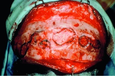
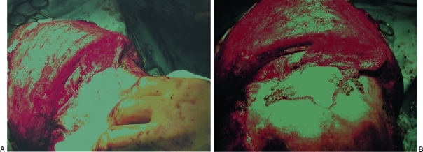
Frontal sinus fracture represents 5 to 12% of all maxillofacial fractures. Because of the anatomic position of the frontal sinus and the enormous amount of force required to create a fracture in this area, these injuries are often devastating and associated with other trauma. Associated injuries include skull base, intracranial, ophthalmologic, and maxillofacial. Complications should be categorized to address these four areas as well as the skin-soft tissue envelope, muscle, and bone. Other variables that should be examined are age of the patient, gender, mechanism of injury, fracture pattern, method of repair, and associated injuries. Management of frontal sinus fractures is so controversial that the indications, timing, method of repair, and surveillance remain disputable among several surgical specialties. The one universal truth that is agreed upon is that all patients undergoing reconstructive surgery of the frontal sinus have a lifelong risk for delayed complications. It is hoped that when patients do experience the first symptoms of a complication, they seek immediate medical attention and avoid potentially life-threatening situations and the need for crippling or disfiguring surgery. The best way to facilitate this is through long-term follow-up and routine surveillance.
Keywords: Complication; acute; chronic; fracture; frontal; sinus.
Figures
Similar articles
-
Frontal sinus fractures: management guidelines.Facial Plast Surg. 2005 Aug;21(3):199-206. doi: 10.1055/s-2005-922860. Facial Plast Surg. 2005. PMID: 16307400
-
A novel classification of frontal bone fractures: The prognostic significance of vertical fracture trajectory and skull base extension.J Plast Reconstr Aesthet Surg. 2015 May;68(5):645-53. doi: 10.1016/j.bjps.2015.02.021. Epub 2015 Feb 26. J Plast Reconstr Aesthet Surg. 2015. PMID: 25778872
-
Fractures of the frontal sinus.Clin Plast Surg. 1989 Jan;16(1):115-23. Clin Plast Surg. 1989. PMID: 2924487
-
Trauma in Facial Plastic Surgery: Frontal Sinus Fractures.Facial Plast Surg Clin North Am. 2017 Nov;25(4):503-511. doi: 10.1016/j.fsc.2017.06.004. Facial Plast Surg Clin North Am. 2017. PMID: 28941504 Review.
-
Endoscopically assisted repair of frontal sinus fracture.J Trauma. 2003 Aug;55(2):378-82. doi: 10.1097/01.TA.0000083333.93868.AB. J Trauma. 2003. PMID: 12913655 Review.
Cited by
-
Management of Frontal Bone Fracture in a Tertiary Neurosurgical Care Center-A Retrospective Study.J Neurosci Rural Pract. 2022 Jan 5;13(1):60-66. doi: 10.1055/s-0041-1740615. eCollection 2022 Jan. J Neurosci Rural Pract. 2022. PMID: 35110921 Free PMC article.
-
Trauma of the midface.GMS Curr Top Otorhinolaryngol Head Neck Surg. 2015 Dec 22;14:Doc06. doi: 10.3205/cto000121. eCollection 2015. GMS Curr Top Otorhinolaryngol Head Neck Surg. 2015. PMID: 26770280 Free PMC article. Review.
-
Preservation of frontal sinus anatomy and outflow tract following frontal trauma with dural defect.Plast Reconstr Surg Glob Open. 2015 Mar 6;3(2):e300. doi: 10.1097/GOX.0000000000000271. eCollection 2015 Feb. Plast Reconstr Surg Glob Open. 2015. PMID: 25750839 Free PMC article.
-
A Case of Penetrating Brain Injury Followed by Delayed Cerebrospinal Fluid Leakage.Korean J Neurotrauma. 2021 Oct 11;17(2):168-173. doi: 10.13004/kjnt.2021.17.e29. eCollection 2021 Oct. Korean J Neurotrauma. 2021. PMID: 34760829 Free PMC article.
-
Frontal Bone Fractures and Frontal Sinus Injuries: Treatment Paradigms.Ann Maxillofac Surg. 2019 Jul-Dec;9(2):261-282. doi: 10.4103/ams.ams_151_19. Ann Maxillofac Surg. 2019. PMID: 31909005 Free PMC article.
References
-
- Manolidis S, Hollier L H., Jr Management of frontal sinus fractures. Plast Reconstr Surg. 2007;120(Suppl 2):32s–48s. - PubMed
-
- Metzinger S E, Guerra A B, Garcia R E. Frontal sinus fractures: management guidelines. Facial Plast Surg. 2005;21:199–206. - PubMed
-
- Xie C, Mehendale N, Barrett D, Bui C J, Metzinger S E. 30-year retrospective review of frontal sinus fractures: the Charity Hospital experience. J Craniomaxillofac Trauma. 2000;6:7–15. discussion 16–18. - PubMed
-
- Strong E B, Pahlavan N, Saito D. Frontal sinus fractures: a 28-year retrospective review. Otolaryngol Head Neck Surg. 2006;135:774–779. - PubMed
-
- Piccolino P, Vetrano S, Mundula P, Di Lella G, Tedaldi M, Poladas G. Frontal bone fractures: new technique of closed reduction. J Craniofac Surg. 2007;18:695–698. - PubMed
LinkOut - more resources
Full Text Sources