

Review of bone substitutes
- PMID: 22110809
- PMCID: PMC3052658
- DOI: 10.1055/s-0029-1224777
Review of bone substitutes
Abstract
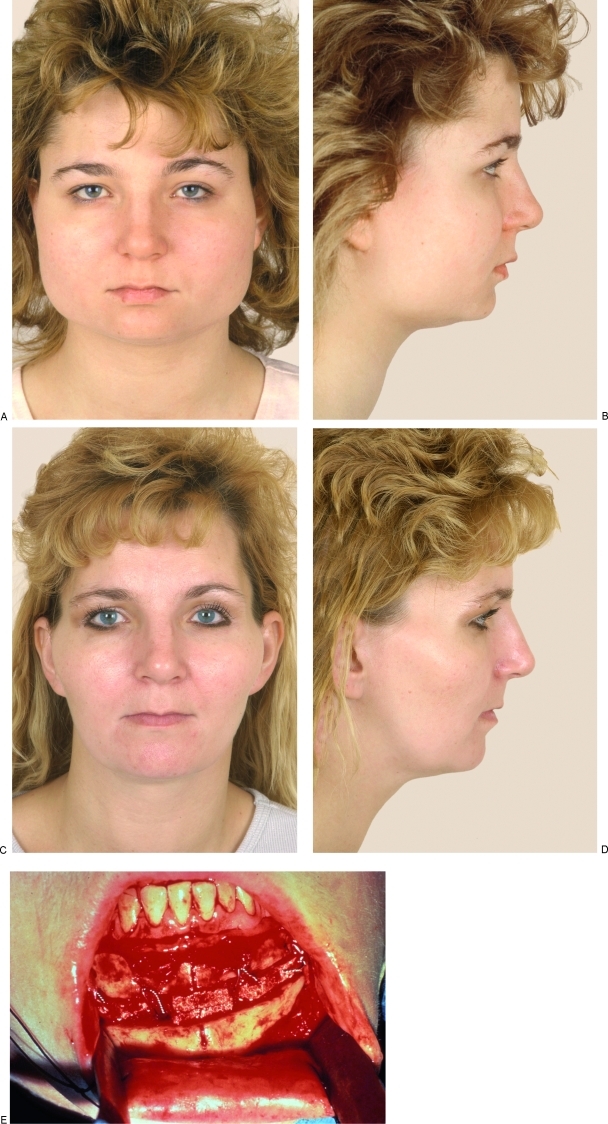
Bone substitutes are being increasingly used in craniofacial surgery and craniomaxillofacial trauma. We will review the history of the biomaterials and describe the ideal characteristics of bone substitutes, with a specific emphasis on craniofacial reconstruction. Some of the most commonly used bone substitutes are discussed in more depth, such as calcium phosphate and hydroxyapatite ceramics and cements, bioactive glass, and polymer products. Areas of active research and future directions include tissue engineering, with an increasing emphasis on bioactivity of the implant.
Keywords: Bone substitutes; biomaterials; calcium phosphate cements; hydroxyapatite; polymethylmethacrylate; porous polyethylene.
Figures


Similar articles
-
Bone substitutes: a review of their characteristics, clinical use, and perspectives for large bone defects management.J Tissue Eng. 2018 Jun 4;9:2041731418776819. doi: 10.1177/2041731418776819. eCollection 2018 Jan-Dec. J Tissue Eng. 2018. PMID: 29899969 Free PMC article. Review.
-
Promoting osteoblast proliferation on polymer bone substitutes with bone-like structure by combining hydroxyapatite and bioactive glass.Mater Sci Eng C Mater Biol Appl. 2019 Mar;96:1-9. doi: 10.1016/j.msec.2018.11.006. Epub 2018 Nov 5. Mater Sci Eng C Mater Biol Appl. 2019. PMID: 30606514
-
Fabrication, Properties and Applications of Dense Hydroxyapatite: A Review.J Funct Biomater. 2015 Dec 21;6(4):1099-140. doi: 10.3390/jfb6041099. J Funct Biomater. 2015. PMID: 26703750 Free PMC article. Review.
-
Multifunctional bioactive glass and glass-ceramic biomaterials with antibacterial properties for repair and regeneration of bone tissue.Acta Biomater. 2017 Sep 1;59:2-11. doi: 10.1016/j.actbio.2017.06.046. Epub 2017 Jul 1. Acta Biomater. 2017. PMID: 28676434 Review.
-
Novel porous hydroxyapatite prepared by combining H2O2 foaming with PU sponge and modified with PLGA and bioactive glass.J Biomater Appl. 2007 Apr;21(4):351-74. doi: 10.1177/0885328206063905. Epub 2006 Mar 16. J Biomater Appl. 2007. PMID: 16543281
Cited by
-
FDA-approved bone grafts and bone graft substitute devices in bone regeneration.Mater Sci Eng C Mater Biol Appl. 2021 Nov;130:112466. doi: 10.1016/j.msec.2021.112466. Epub 2021 Sep 29. Mater Sci Eng C Mater Biol Appl. 2021. PMID: 34702541 Free PMC article. Review.
-
Cellular Response of Human Osteoblasts to Different Presentations of Deproteinized Bovine Bone.Materials (Basel). 2022 Jan 27;15(3):999. doi: 10.3390/ma15030999. Materials (Basel). 2022. PMID: 35160947 Free PMC article.
-
The Biological Evaluation of Conventional and Nano-Hydroxyapatite-Silica Glass Ionomer Cement on Dental Pulp Stem Cells: A Comparative Study.Contemp Clin Dent. 2019 Apr-Jun;10(2):324-332. doi: 10.4103/ccd.ccd_581_18. Contemp Clin Dent. 2019. PMID: 32308298 Free PMC article.
-
Incorporating strontium enriched amorphous calcium phosphate granules in collagen/collagen-magnesium-hydroxyapatite osteochondral scaffolds improves subchondral bone repair.Mater Today Bio. 2024 Jan 20;25:100959. doi: 10.1016/j.mtbio.2024.100959. eCollection 2024 Apr. Mater Today Bio. 2024. PMID: 38327976 Free PMC article.
-
Bone Morphogenetic Protein as Bone Additive around Dental Implant and its Impact on Osseointegration: a Systematic Review.J Dent (Shiraz). 2022 Sep;23(2 Suppl):336-348. doi: 10.30476/DENTJODS.2021.90931.1536. J Dent (Shiraz). 2022. PMID: 36588970 Free PMC article.
References
-
- Urist M R, O'Conner B T, Burwell R G. Bone Graft, Derivatives and Substitutes. Cambridge: Butterworth-Heinemann; 1994.
-
- Flati G, Di Stanislao C. Chirurgia nella preistoria. Parte I. Provincia Med Aquila. 2004;2:8–11.
-
- Donati D, Zolezzi C, Tomba P, Viganò A. Bone grafting: historical and conceptual review, starting with an old manuscript by Vittorio Putti. Acta Orthop. 2007;78:19–25. - PubMed
-
- De Boer H H. The history of bone grafts. Clin Orthop Relat Res. 1988;226:292–298. - PubMed
-
- von Walter P. J Chir und Augen-Heilkunde. 1821;2:571.
LinkOut - more resources
Full Text Sources
Other Literature Sources
Medical