

Traumatic superior orbital fissure syndrome: current management
- PMID: 22110813
- PMCID: PMC3052661
- DOI: 10.1055/s-0030-1249369
Traumatic superior orbital fissure syndrome: current management
Abstract
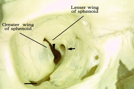
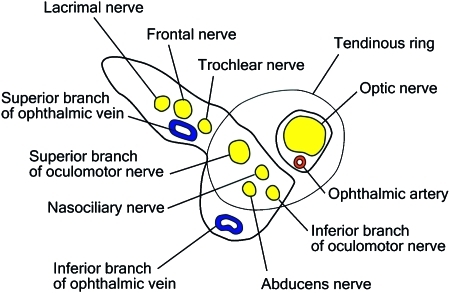
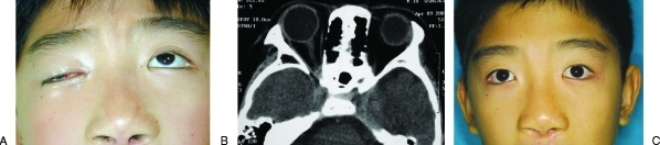
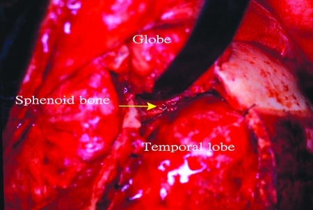
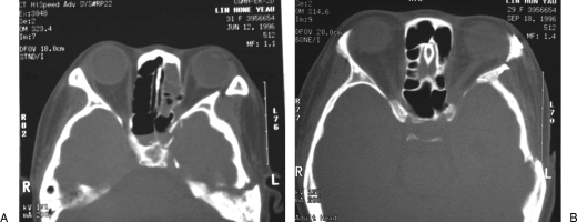
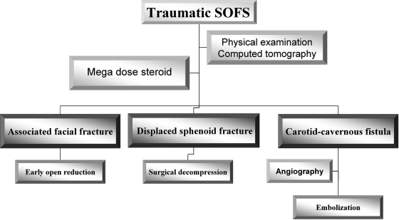
Traumatic superior orbital fissure syndrome is an uncommon complication of craniomaxillofacial trauma with an incidence of less than 1%. The syndrome is characterized by ophthalmoplegia, ptosis, proptosis of eye, dilation and fixation of the pupil, and anesthesia of the upper eyelid and forehead. This article describes a detailed anatomy of the superior orbital fissure as it related to pathophysiology and clinical findings. Etiology and diagnosis are established after detailed physical and radiographic examination. On the basis of our clinical experience in the management of superior orbital fissure syndrome and from the data reported previously in the literature, an algorithm for treatment of traumatic superior orbital fissure syndrome including use of steroid, surgical decompression of superior orbital fissure, and reduction of concomitant facial fracture is presented and its rationale discussed.
Keywords: Superior orbital fissure; cranial nerve; optic nerve; orbital apex syndrome; steroid.
Figures
References
-
- Banks P. The superior orbital fissure syndrome. Oral Surg Oral Med Oral Pathol. 1967;24:455–458. - PubMed
-
- Lakke J P. Superior orbital fissure syndrome. Report of a case caused by local pachymeningitis. Arch Neurol. 1962;7:289–300. - PubMed
-
- Govsa F, Kayalioglu G, Erturk M, Ozgur T. The superior orbital fissure and its contents. Surg Radiol Anat. 1999;21:181–185. - PubMed
-
- Karakaş P, Bozkir M G, Oguz O. Morphometric measurements from various reference points in the orbit of male Caucasians. Surg Radiol Anat. 2003;24:358–362. - PubMed
-
- Morard M, Tcherekayev V, de Tribolet N. The superior orbital fissure: a microanatomical study. Neurosurgery. 1994;35:1087–1093. - PubMed
LinkOut - more resources
Full Text Sources
Miscellaneous