. 2010 Jun;3(2):105-13.
doi: 10.1055/s-0030-1254383.

Management of temporal bone trauma

Management of temporal bone trauma

Alpen Patel et al. Craniomaxillofac Trauma Reconstr. 2010 Jun.

Abstract

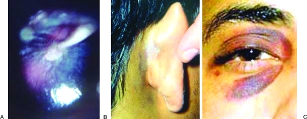
The temporal bones are paired structures located on the lateral aspects of the skull and contribute to the skull base. Trauma is usually the result of blunt head injury and can result in damage to the brain and meninges, the middle and internal ear, and the facial nerve. Complications can include intracranial hemorrhage, cerebral contusion, CSF leak and meningitis, hearing loss, vertigo, and facial paralysis. To prevent these complications, diagnosis followed by appropriate medical and surgical management is critical. Diagnosis relies primarily on physical signs and symptoms as well as radiographic imaging. Emergent intervention is required in situations involving herniation of the brain into the middle ear cavity or hemorrhage of the intratemporal carotid artery. Patients with declining facial nerve function are candidates for early surgical intervention. Conductive hearing loss can be corrected surgically as an elective procedure, while sensorineural hearing loss carries a poor prognosis, regardless of management approach. Children generally recover from temporal bone trauma with fewer complications than adults and experience a markedly lower incidence of facial nerve paralysis.

Keywords: Temporal bone; management; trauma.


Figures

Figure 1
Figure 1
Diagram of the paired temporal bones (shaded) in relation to the human skull. Anterior view (A), right lateral view (B), inferior view (C), and interior view of the skull base (D).
Figure 2
Figure 2
Diagram of the left temporal bone from a lateral view. The squamous, styloid, and mastoid portions are labeled. The tympanic portion lines the external auditory meatus and the petrous portion is an interior structure and is not visible from a lateral view.
Figure 3
Figure 3
Schematic diagram of the right external, middle, and internal ear. Sound travels through the external acoustic meatus causing the tympanic membrane to vibrate. The ossicles of the middle ear (malleus, incus, and stapes) amplify and transmit these vibrations through the oval window to the cochlea, an internal ear structure. Vibrations are then translated into neural impulses and carried to the brain via the vestibulocochlear nerve (CN VIII). CN VIII also transmits information from the vestibular apparatus (balance organ) of the internal ear, which includes the three semicircular canals, and the utricle and saccule (not shown).
Figure 4
Figure 4
Cerebrospinal fluid (CSF) leaking from the ear (otorrhea) indicates that a temporal bone fracture has occurred and that the tympanic membrane has been disrupted. Leakage from the nose (rhinorrhea) is also indicative of a basilar skull fracture.
Figure 5
Figure 5
A presumptive diagnosis of temporal bone fracture can be made with the presence of three physical findings. Hemotympanum (A) is a collection of blood in the middle ear and gives the tympanic membrane and reddish-blue hue when visualized externally. Postauricular ecchymosis, or Battle's sign (B), is an arch-shaped bruise behind the external ear. Periorbital ecchymosis, or Raccoon sign (C), is circular bruising around the eye.
Figure 6
Figure 6
Diagram of common fracture patterns of the temporal bone. Transverse fractures (A) result most often from a blow to the back of the head. The fracture extends from the jugular foramen through the petrous pyramid to the foramen spinosum and foramen lacerum. Longitudinal fractures (B) result most often from a blow to the side of the head. The fracture extends from the squamous portion of the temporal bone to the carotid and jugular foramina.
Figure 7
Figure 7
Axial computed tomography (CT) scan of the showing both a longitudinal fracture (small arrowhead) and a transverse fracture (large arrowhead) of the left temporal bone.
Figure 8
Figure 8
Diagram of the facial nerve and other middle ear structures as exposed in a transmastoid approach. This approach is suitable for patients whose nerve injury lies distal to the geniculate ganglion. The incus can be temporarily removed to facilitate this approach.
Figure 9
Figure 9
Diagram of the facial nerve as exposed in a middle cranial fossa approach. Careful dissection is essential to avoid the superior semicircular canal and the basal turn of the cochlea.
Figure 10
Figure 10
Diagram of the facial nerve as exposed in a translabyrinthine approach. When combined with a transmastoid approach, the entire intratemporal course of the facial nerve can be visualized.

References

    1. Gunlock M G, Gentry L R. Anatomy of the temporal bone. Neuroimaging Clin N Am. 1998;8(1):195–209. - PubMed
    1. Proctor B. The anatomy of the facial nerve. Otolaryngol Clin North Am. 1991;24(3):479–504. - PubMed
    1. Hasso A N, Ledington J A. Traumatic injuries of the temporal bone. Otolaryngol Clin North Am. 1988;21(2):295–316. - PubMed
    1. Wiet R J, Valvassori G E, Kotsanis C A, et al. Temporal bone fractures: state of the art review. Am J Otol. 1985;6(3):207–215. - PubMed
    1. Shindo M L, Fetterman B L, Shih L, Maceri D R, Rice D H. Gunshot wounds of the temporal bone: a rational approach to evaluation and management. Otolaryngol Head Neck Surg. 1995;112(4):533–539. - PubMed

LinkOut - more resources