

Sinus preservation management for frontal sinus fractures in the endoscopic sinus surgery era: a systematic review
- PMID: 22110830
- PMCID: PMC3052679
- DOI: 10.1055/s-0030-1262957
Sinus preservation management for frontal sinus fractures in the endoscopic sinus surgery era: a systematic review
Abstract
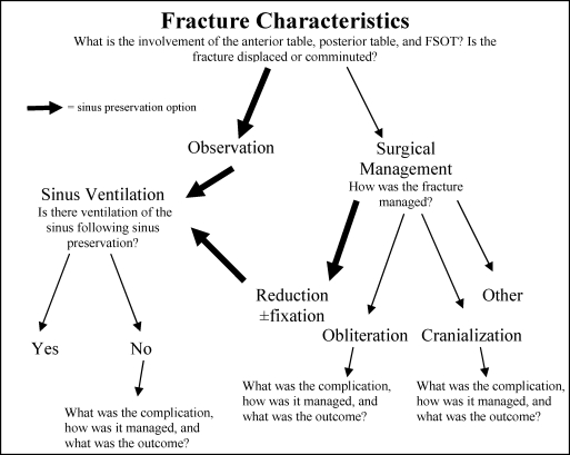
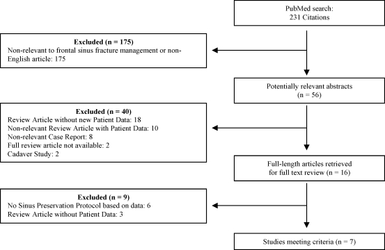
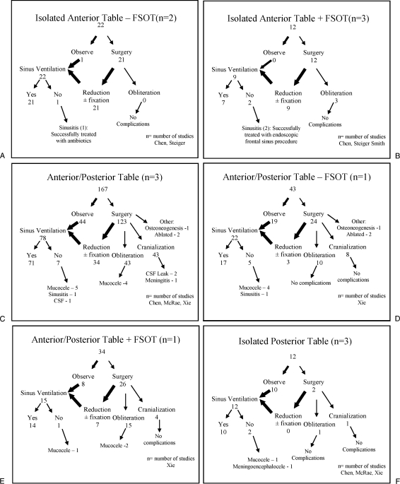
We systematically reviewed the existing literature supporting the efficacy and safety of sinus preservation management for frontal sinus fractures in the modern era of endoscopic frontal sinus surgery. A systematic review of the English literature for the targeted objective was conducted using the PubMed database between January 1995 and August 2008. The PubMed database was queried using two major search terms of frontal sinus fracture or frontal sinus injury along with manual review of citations within bibliographies. Citations acquired from the primary search were filtered and relevant abstracts were identified that merited full review. Articles were identified that included any cohort of patients with frontal sinus fractures involving the frontal sinus outflow tract or posterior wall with sinus preservation management. A total of 231 citations were generated, and 56 abstracts were identified as potentially relevant articles. Sixteen articles merited full review, with seven articles meeting inclusion criteria for sinus preservation. There were 515 total patients in the studies with 350 patients managed with frontal sinus preservation. Similar short-term complications and effectiveness were found between fractures managed with sinus preservation and those with traditional management. Sinus preservation appears to be a safe and effective management strategy for select frontal sinus fractures. More transparent reporting of management strategies for individual cases or cohorts is needed. A standardized algorithm and categorization framework for future studies are proposed. Longer-term follow-up and larger prospective studies are necessary to assess the safety and efficacy of sinus preservation protocols.
Keywords: Frontal sinus trauma; endoscopic sinus surgery; frontal sinus outflow tract; osteoplastic obliteration; sinus cranialization; sinus preservation.
Figures
Similar articles
-
Endoscopic management of the frontal recess in frontal sinus fractures: a shift in the paradigm?Laryngoscope. 2002 May;112(5):784-90. doi: 10.1097/00005537-200205000-00004. Laryngoscope. 2002. PMID: 12150607
-
Frontal sinus fractures with suspected outflow tract obstruction: a new approach for sinus preservation.J Craniomaxillofac Surg. 2015 Jan;43(1):1-6. doi: 10.1016/j.jcms.2014.09.013. Epub 2014 Oct 29. J Craniomaxillofac Surg. 2015. PMID: 25458344
-
Frontal Sinus Fractures: Evolving Clinical Considerations and Surgical Approaches.Craniomaxillofac Trauma Reconstr. 2019 Jun;12(2):85-94. doi: 10.1055/s-0039-1678660. Epub 2019 Feb 4. Craniomaxillofac Trauma Reconstr. 2019. PMID: 31073357 Free PMC article. Review.
-
Frontal sinus fracture management: a systematic review and meta-analysis.Int J Oral Maxillofac Surg. 2021 Jan;50(1):75-82. doi: 10.1016/j.ijom.2020.06.004. Epub 2020 Aug 27. Int J Oral Maxillofac Surg. 2021. PMID: 32861554
-
Is there still a role for cranialization in modern sinus surgery?Curr Opin Otolaryngol Head Neck Surg. 2021 Feb 1;29(1):53-58. doi: 10.1097/MOO.0000000000000691. Curr Opin Otolaryngol Head Neck Surg. 2021. PMID: 33278134 Review.
Cited by
-
Endoscopic management of frontal sinus diseases after frontal craniotomy: a case series and review of the literature.Eur Arch Otorhinolaryngol. 2021 Apr;278(4):1035-1045. doi: 10.1007/s00405-020-06335-7. Epub 2020 Sep 3. Eur Arch Otorhinolaryngol. 2021. PMID: 32880737 Review.
-
A Naso-Orbito-Ethmoid (NOE) Fracture Associated with Bilateral Anterior and Posterior Frontal Sinus Wall Fractures Caused by a Horse Kick-Case Report and Short Literature Review.Medicina (Kaunas). 2019 Nov 9;55(11):731. doi: 10.3390/medicina55110731. Medicina (Kaunas). 2019. PMID: 31717521 Free PMC article.
-
Secondary Reconstruction of Frontal Sinus Fracture.Arch Craniofac Surg. 2016 Sep;17(3):103-110. doi: 10.7181/acfs.2016.17.3.103. Epub 2016 Sep 23. Arch Craniofac Surg. 2016. PMID: 28913266 Free PMC article. Review.
-
Trauma of the midface.GMS Curr Top Otorhinolaryngol Head Neck Surg. 2015 Dec 22;14:Doc06. doi: 10.3205/cto000121. eCollection 2015. GMS Curr Top Otorhinolaryngol Head Neck Surg. 2015. PMID: 26770280 Free PMC article. Review.
-
Evaluation of a Minimally Disruptive Treatment Protocol for Frontal Sinus Fractures.JAMA Facial Plast Surg. 2017 May 1;19(3):225-231. doi: 10.1001/jamafacial.2016.1769. JAMA Facial Plast Surg. 2017. PMID: 28152148 Free PMC article.
References
-
- Bell R B, Dierks E J, Brar P, Potter J K, Potter B E. A protocol for the management of frontal sinus fractures emphasizing sinus preservation. J Oral Maxillofac Surg. 2007;65:825–839. - PubMed
-
- Rohrich R J, Hollier L H. Management of frontal sinus fractures. Changing concepts. Clin Plast Surg. 1992;19:219–232. - PubMed
-
- McRae M, Momeni R, Narayan D. Frontal sinus fractures: a review of trends, diagnosis, treatment, and outcomes at a level 1 trauma center in Connecticut. Conn Med. 2008;72:133–138. - PubMed
-
- Tiwari P, Higuera S, Thornton J, Hollier L H. The management of frontal sinus fractures. J Oral Maxillofac Surg. 2005;63:1354–1360. - PubMed
-
- Manolidis S, Hollier L H., Jr Management of frontal sinus fractures. Plast Reconstr Surg. 2007;120(7 Suppl 2):32S–48S. - PubMed
LinkOut - more resources
Full Text Sources
Other Literature Sources