

Primary intestinal lymphangiectasia diagnosed by capsule endoscopy and double balloon enteroscopy
- PMID: 22110841
- PMCID: PMC3221958
- DOI: 10.4253/wjge.v3.i11.235
Primary intestinal lymphangiectasia diagnosed by capsule endoscopy and double balloon enteroscopy
Abstract
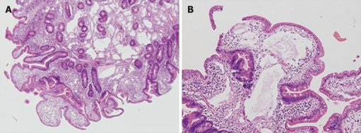
Primary intestinal lymphangiectasia (PIL) is a rare disorder characterized by dilated intestinal lymphatics and the development of protein-losing enteropathy. Patients with PIL develop hypoalbuminemia, hypocalcemia, lymphopenia and hypogammaglobulinemia, and present with bilateral lower limb edema, fatigue, abdominal pain and diarrhea. Endoscopy reveals diffusely elongated, circumferential and polypoid mucosae covered with whitish enlarged villi, all of which indicate intestinal lymphangiectasia. Diagnosis is confirmed by characteristic tissue pathology, which includes dilated intestinal lymphatics with diffusely swollen mucosa and enlarged villi. The prevalence of PIL has increased since the introduction of capsule endoscopy. The etiology and prevalence of PIL remain unknown. Some studies have reported that several genes and regulatory molecules for lymphangiogenesis are related to PIL. We report the case of a patient with PIL involving the entire small bowel that was confirmed by capsule endoscopy and double-balloon enteroscopy-guided tissue pathology who carried a deletion on chromosome 4q25. The relationship between this deletion on chromosome 4 and PIL remains to be investigated.
Keywords: Capsule endoscopy; Chromosome 4q25; Chromosome deletion; Double balloon enteroscopy; Primary intestinal lymphangiectasia.
Figures


Similar articles
-
Case report of primary intestinal lymphangiectasia diagnosed in an octogenarian by ileal intubation and by push enteroscopy after missed diagnosis by standard colonoscopy and EGD.Medicine (Baltimore). 2018 Jan;97(3):e9649. doi: 10.1097/MD.0000000000009649. Medicine (Baltimore). 2018. PMID: 29505002 Free PMC article.
-
Primary intestinal lymphangiectasia (Waldmann's disease).Orphanet J Rare Dis. 2008 Feb 22;3:5. doi: 10.1186/1750-1172-3-5. Orphanet J Rare Dis. 2008. PMID: 18294365 Free PMC article. Review.
-
Primary intestinal lymphangiectasia in an adult patient: A case report and review of literature.World J Gastroenterol. 2020 Dec 28;26(48):7707-7718. doi: 10.3748/wjg.v26.i48.7707. Epub 2020 Dec 8. World J Gastroenterol. 2020. PMID: 33505146 Free PMC article. Review.
-
Ileal polypoid lymphangiectasia bleeding diagnosed and treated by double balloon enteroscopy.World J Gastroenterol. 2013 Dec 7;19(45):8440-4. doi: 10.3748/wjg.v19.i45.8440. World J Gastroenterol. 2013. PMID: 24363538 Free PMC article. Review.
-
Elderly Onset Primary Intestinal Lymphangiectasia-A Rare Case.JGH Open. 2025 Jan 22;9(1):e70102. doi: 10.1002/jgh3.70102. eCollection 2025 Jan. JGH Open. 2025. PMID: 39850090 Free PMC article.
Cited by
-
Primary intestinal lymphangiectasia diagnosed by video capsule endoscopy in a patient with immunodeficiency presenting with Morganella morganii bacteraemia.BMJ Case Rep. 2020 Sep 13;13(9):e235898. doi: 10.1136/bcr-2020-235898. BMJ Case Rep. 2020. PMID: 32928820 Free PMC article.
-
Primary intestinal lymphangiectasia in an elderly female patient: A case report on a rare cause of secondary immunodeficiency.Medicine (Baltimore). 2017 Aug;96(31):e7729. doi: 10.1097/MD.0000000000007729. Medicine (Baltimore). 2017. PMID: 28767614 Free PMC article.
-
An unusual cause of ankle swelling.Endosc Int Open. 2014 Dec;2(4):E262-4. doi: 10.1055/s-0034-1377382. Epub 2014 Aug 1. Endosc Int Open. 2014. PMID: 26135105 Free PMC article. No abstract available.
-
Small intestinal mucosal abnormalities using video capsule endoscopy in intestinal lymphangiectasia.Orphanet J Rare Dis. 2023 Oct 2;18(1):308. doi: 10.1186/s13023-023-02914-z. Orphanet J Rare Dis. 2023. PMID: 37784188 Free PMC article.
-
Internal frontier: the pathophysiology of the small intestine.World J Gastroenterol. 2013 Jan 14;19(2):161-4. doi: 10.3748/wjg.v19.i2.161. World J Gastroenterol. 2013. PMID: 23345938 Free PMC article.
References
-
- WALDMANN TA, STEINFELD JL, DUTCHER TF, DAVIDSON JD, GORDON RS. The role of the gastrointestinal system in "idiopathic hypoproteinemia". Gastroenterology. 1961;41:197–207. - PubMed
-
- Salomons HA, Kramer P, Nikulasson S, Schroy PC. Endoscopic features of long-standing primary intestinal lymphangiectasia. Gastrointest Endosc. 1995;41:516–518. - PubMed
-
- Lenzhofer R, Lindner M, Moser A, Berger J, Schuschnigg C, Thurner J. Acute jejunal ileus in intestinal lymphangiectasia. Clin Investig. 1993;71:568–571. - PubMed
-
- Dierselhuis MP, Boelens JJ, Versteegh FG, Weemaes C, Wulffraat NM. Recurrent and opportunistic infections in children with primary intestinal lymphangiectasia. J Pediatr Gastroenterol Nutr. 2007;44:382–385. - PubMed
LinkOut - more resources
Full Text Sources
