

Necrobiosis and T-lymphocyte infiltration in retrieved aseptically loosened metal-on-polyethylene arthroplasties
- PMID: 22112191
- PMCID: PMC3242957
- DOI: 10.3109/17453674.2011.625534
Necrobiosis and T-lymphocyte infiltration in retrieved aseptically loosened metal-on-polyethylene arthroplasties
Abstract
Background and purpose: Soft tissue necrobiosis and T-lymphocyte infiltration within the periprosthetic soft tissue have been linked to a suggested hypersensitivity reaction of the delayed-type following the metal-on-metal arthroplasty. While we observed both synovial necrobiosis and lymphocyte infiltrates in synovial tissues with failed metal-on-polyethylene prostheses, we hypothesized that both findings are unspecific for metal-on-metal bearing coupes. Thus, we wished to quantify the extent of necrobiosis and the amount of T-lymphocyte infiltration in aseptically loosened metal-on-polyethylene prostheses.
Materials and methods: We analyzed 28 consecutive synovial biopsy specimens obtained at revision surgery of aseptically loosened metal-on-polyethylene prostheses (19 hips and 9 knees) and quantified both the extent of necrobiosis vertically and the density of CD3+, CD4+, and CD8+ lymphocytes within the joint capsular tissue. We excluded patients with inflammatory skeletal disease or with a history of metal hypersensitivity.
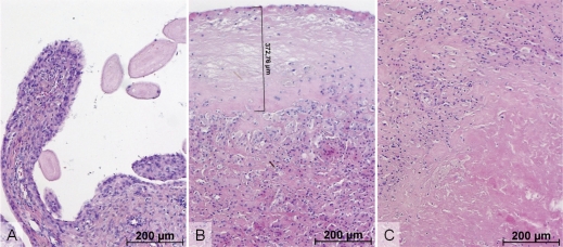
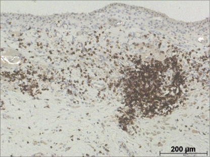
Results: We found necrobiosis in 23 of 28 cases and it was most often connected with the superficial portions of the synovium. Necrobiosis of deeper tissues was seen in 8 specimens and it was strongly associated with superficial necrobiosis. While CD3+ lymphocytes were detected in each biopsy, 4 cases with more than 300 CD3+ lymphocytes were identified in the group of 26 cases that presented with more than 100 CD3+ lymphocytes within one high-power field. 16 cases with more than 100 CD3+ lymphocytes also showed concomitant superficial necrobiosis of the synovium. In the inflammatory infiltration of periprosthetic synovium, CD8+ lymphocytes predominated over CD4+ cells.
Interpretation: Synovial necrobiosis and infiltration of T-lymphocytes are common findings in tissues around aseptically loosened metal-on-polyethylene arthroplasty in patients without a clinically suspected metal hypersensitivity reaction. Thus, neither necrobiosis nor infiltration of T-lymphocytes should be considered to be specific for failed metal-on-metal bearings or metal hypersensitivity reaction.
Figures
References
-
- Doorn PF, Mirra JM, Campbell PA, Amstutz HC. Tissue reaction to metal on metal total hip prostheses. Clin Orthop (Suppl) 1996;329:S187–205. - PubMed
-
- Fang CS, Harvie P, Gibbons CL, Whitwell D, Athanasou NA, Ostlere S. The imaging spectrum of peri-articular inflammatory masses following metal-on-metal hip resurfacing. Skeletal Radiol. 2008;37(8):715–22. - PubMed
MeSH terms
Substances
LinkOut - more resources
Full Text Sources
Medical
Research Materials